SANTA CLARA, Calif., November 13, 2023 (Newswire.com)
Zeto, Inc., an innovative EEG brain monitoring company, is excited to make two significant announcements as the company continues to expand the boundaries of brain monitoring.
Zeto is proud to announce its inaugural participation at the Neuroscience 2023 conference hosted by the Society for Neuroscience. Taking place from November 11-15, 2023, at the Walter E. Washington Convention Center in Washington, D.C., this event will bring together over 30,000 scientists, researchers, and clinicians making it the largest conference in neuroscience.
“At Zeto, we’ve consistently delivered substantial value to our clinical partners in hospitals and private practices. Now, we’re excited to bring the convenience of Zeto EEG to the forefront of your clinical research journey. The Society for Neuroscience conference is a remarkable opportunity for us to connect with the global scientific community and facilitate the use of EEG for brain monitoring,” said Florian Strelzyk, PhD, Chief Sales Officer at Zeto. “Zeto understands your need for ease of use, comfort, and a refined experience with EEG technology. We believe our products will unlock a world of new opportunities, enabling EEG researchers to step out of the lab and into research environments closely aligned with their interests. We invite attendees to visit our booth #2430 in the Exhibitor Hall to discover and experience our innovative EEG solutions for clinical trials and research.”
Zeto reaffirms its commitment to increasing accessibility to brain monitoring by sponsoring up to three EEG clinical trials in 2024. These trials aim to support advanced clinical research and bring innovative applications closer to full real-world implementation.
Each sponsored clinical trial will enjoy the following benefits:
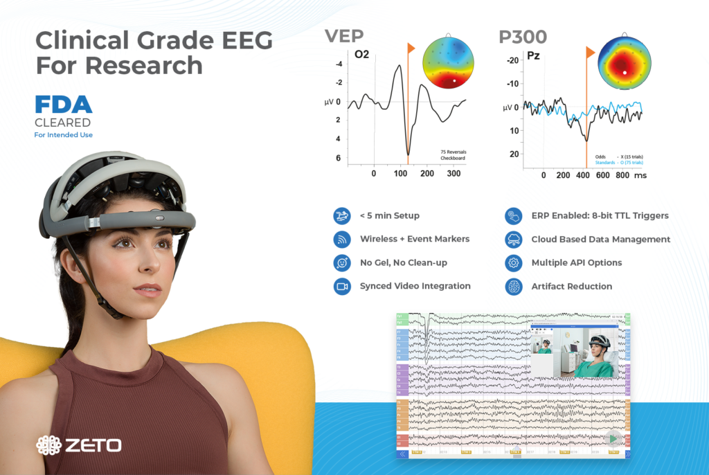
WR19 EEG System: Each sponsored clinical trial will benefit from Zeto’s EEG device, an FDA-cleared, wireless, portable, dry-electrode, full 10-20 montage EEG headset with high-quality data collection and a fast and comfortable 5-minute setup.
Cloud Access: Access to HIPAA-compliant, cyber-security cleared cloud platform for efficient data storage, remote access, EDF export, and real-time LSL data streaming.
Disposable Electrodes: Zeto will provide disposable electrodes for up to 100 EEG recordings in each clinical trial. These electrodes are designed for comfort and ease of use, enhancing the participant experience.
TTL-based Triggering for ERP Recordings: If required, the Zeto system will include an additional component for real-time TTL-based triggering for evoked potentials, along with additional stimulus presentation software and hardware.
Training and Support: To ensure a smooth and successful implementation of Zeto’s EEG solutions, the company provides training and ongoing support to clinical trial teams.
To participate in this opportunity, interested research teams are invited to submit a 2-page proposal or project pitch by March 31st, 2024. Further details and submission guidelines can be found on the Zeto Clinical Trials Sponsorship webpage.
Zeto is dedicated to enhancing the accessibility of brain monitoring. For more information about Zeto, please visit Zeto’s website.
The American Heart Association (AHA) recommends prompt electroencephalography (EEG) neuroprognostication for post-cardiac arrest patients in their 2020 guidelines on cardiopulmonary resuscitation (CPR) and emergency cardiovascular care (ECC). The prompt use of EEG in post-cardiac arrest patients is important because it allows for early identification of brain injury and can guide decisions about the continuation of life-sustaining treatment.
Hypoxic-ischemic brain injury is a leading cause of morbidity and mortality in survivors of hospital cardiac arrest.1 Sadly, most of the post-resuscitation deaths are caused by the active withdrawal of life-sustaining treatment. The decision to actively remove life-sustaining treatment is made when a poor neurological outcome is expected. For this reason, it is critical to perform accurate neuroprognostication, as recommended by the American Heart Association (AHA) in their 2020 guidelines on cardiopulmonary resuscitation (CPR) and emergency cardiovascular care (ECC).2
Proper post-cardiac arrest neuroprognostication, or EEG after cardiac arrest, is essential to distinguish those who may achieve a meaningful neurological recovery from those who will inevitably have a poor neurological outcome.2,3
Who Should Receive Neuroprognostication After Cardiac Arrest?
The AHA recommends multimodal neuroprognostication on all patients who remain comatose after cardiac arrest (Level 1 recommendation).2 Multimodal neuroprognostication includes EEG, MRI, quantitative pupillometry, and serum neuron-specific enolase, among others. In addition to neuroprognostication, EEG testing is recommended to identify seizures and, if necessary, provide treatment.
Nonconvulsive seizures, for example, are common after cardiac arrest, and cannot be reliably detected without EEG.4 The American Academy of Neurology also provides data on its importance. Also, EEG after cardiac arrest should be used to rule out underlying ictal activity in cardiac arrest survivors with status myoclonus. The 2020 Emergency Cardiovascular Care Science with Treatment Recommendations (CoSTR) advises seizures to be treated when diagnosed in cardiac arrest patients with return of spontaneous circulation (ROSC).5
When Should Neuroprognostication After Cardiac Arrest Start?
Importantly, prognostic assessments should not be started too early. If they are administered too soon after the cardiac arrest and during initial post-resuscitation care, the results may appear worse than they actually are because of medications, or acute post-injury changes.6 Perhaps surprisingly, clinical prognostic testing such as pupillary light reflex should not be used for neuroprognostication until at least 5 days after ROSC (return of spontaneous circulation) in patients treated with targeted temperature management (TTM), in order for such testing to have prognostic significance.
Testing should not begin until the patient has been normothermic for at least 72 hours.6-8 Imaging or EEG to detect status myoclonus may begin as early as 24 hours after ROSC; two studies including 347 patients, showed the presence of status myoclonus within 72 hours of ROSC predicted poor neurological outcome with specificity of 97% to 100%.9,10 However, postanoxic status epilepticus may not manifest until 72 hours or more after ROSC and sedative drug dosages have been reduced, so waiting for neurological prognosis assessment is necessary.
How to Use EEG for Neuroprognostication After ROSC
The 2020 American Heart Association (AHA) Guidelines for Cardiopulmonary Resuscitation (CPR) and Emergency Cardiovascular Care (ECC) recommend the use of EEG after cardiac arrest in patients who remain in a coma after ROSC for the purposes of neuroprognostication.2
Findings that are consistent with poor outcomes include postanoxic status epilepticus and/or burst suppression 72 hours or more after ROSC.2 Another potentially useful electrodiagnostic test is the somatosensory evoked potential (SSEP).
SSEP testing is conducted by stimulating the median nerve and looking for a resulting cortical N20 wave. N20 SSEP waves that are absent bilaterally correlate with poor prognosis.2 Likewise, rhythmic periodic discharges on EEG are also consistent with poor prognosis.
Importantly, the AHA notes that the absence of EEG reactivity within 72 hours after cardiac arrest should not be used as the sole determinant of poor neurological outcomes. A lack of EEG reactivity during this time does not necessarily predict a poor neurological outcome.
Obtaining EEG after ROSC
Conventional EEG in the Intensive Care Unit is Critical Care and Cumbersome
While conventional EEG is an acceptable means to obtain EEG in the ICU, it is impractical. Post-cardiac arrest patients who need post-cardiac arrest care in the ICU will, as standard care, be intubated, and have central venous and possibly arterial lines, and intracardiac devices.
Conventional EEG after cardiac arrest is challenging. Attempting to obtain EEG signals through a dozen wires in a critical care setting without quality-limiting artifacts is challenging. Indeed, providing continuous conventional EEG in the ICU is a literal barrier to care for ICU or neurocritical care nurses and staff.
Zeto EEG – Wireless Full-Montage Monitoring For Use on Coma Patients After ROSC
According to the AHA, proper neuroprognostication could prevent withdrawal of life support as it will indicate the accurate neurologic prognosis of a patient who has a chance at successful neurological recovery. Thus, the AHA recommends performing multimodal neurologic prognostication including EEG in all patients who remain in a coma after ROSC following cardiac arrest.
Zeto has developed a full montage, 19-channel (10-20 system) wireless headset with dry electrodes for rapid EEG monitoring. The benefits of Zeto’s electrodes include no skin preparation, no cleanup, and no gel or paste residue. The electrodes are single-use and soft.
The Zeto EEG device collects high-quality EEG recordings and transmits them wirelessly to the cloud for remote viewing and interpretation. ZETO’s EEG platform also offers an FDA-cleared seizure detection and trending algorithm developed by Encevis.
The Zeto EEG headset can be placed after a short training, and the setup takes only 5 minutes – which is crucial for patients in a coma. Once placed, the Zeto headset provides continuous EEG monitoring for up to 5-6 hours.
Overall, prompt EEG for post-cardiac arrest patients is a vital aspect of proper neuroprognostication and plays an important role in determining prognosis in patients and guiding treatment decisions. It is important for healthcare providers, especially intensive care medical providers to be aware of the AHA guidelines and incorporate EEG into their standard care management of post-cardiac arrest patients.
When called upon to perform the difficult and highly consequential task of neuroprognostication, choose Zeto for EEG in the ICU.
References
Witten L, Gardner R, Holmberg MJ, et al. Reasons for death in patients successfully resuscitated from out-of-hospital and in-hospital cardiac arrest. Resuscitation. 2019;136:93-99. PMID:30710595 doi:10.1016/j.resuscitation.2019.01.031
Panchal AR, Bartos JA, Cabanas JG, et al. Part 3: Adult Basic and Advanced Life Support: 2020 American Heart Association Guidelines for Cardiopulmonary Resuscitation and Emergency Cardiovascular Care. Circulation. 2020;142(16_suppl_2):S366-S468. PMID:33081529 doi:10.1161/CIR.0000000000000916
Geocadin RG, Callaway CW, Fink EL, et al. Standards for Studies of Neurological Prognostication in Comatose Survivors of Cardiac Arrest: A Scientific Statement From the American Heart Association. Circulation. 2019;140(9):e517-e542. PMID:31291775 doi:10.1161/CIR.0000000000000702
Freund B, Kaplan PW. Myoclonus After Cardiac Arrest: Where Do We Go From Here? Epilepsy Curr. 2017 Sep-Oct;17(5):265-272. doi: 10.5698/1535-7597.17.5.265. PMID: 29225535; PMCID: PMC5716491.
Ryoo SM, Jeon SB, Sohn CH, et al. Predicting Outcome With Diffusion-Weighted Imaging in Cardiac Arrest Patients Receiving Hypothermia Therapy: Multicenter Retrospective Cohort Study. Crit Care Med. 2015;43(11):2370-2377. PMID:26284621 doi:10.1097/CCM.0000000000001263
Samaniego EA, Mlynash M, Caulfield AF, Eyngorn I, Wijman CA. Sedation confounds outcome prediction in cardiac arrest survivors treated with hypothermia. Neurocrit Care. 2011;15(1):113-119. PMID:20680517 doi:10.1007/s12028-010-9412-8
Berg KM, Soar J, Andersen LW, et al. Adult Advanced Life Support: International Consensus on Cardiopulmonary Resuscitation and Emergency Cardiovascular Care Science With Treatment Recommendations. Resuscitation. 2020. PMID:33098922 doi:10.1016/j.resuscitation.2020.09.012
Callaway CW, Donnino MW, Fink EL, et al. Part 8: Post-Cardiac Arrest Care: 2015 American Heart Association Guidelines Update for Cardiopulmonary Resuscitation and Emergency Cardiovascular Care. Circulation. 2015;132(18 Suppl 2):S465-482. PMID:26472996 doi:10.1161/CIR.0000000000000262
Ruknuddeen MI, Ramadoss R, Rajajee V, Grzeskowiak LE, Rajagopalan RE. Early clinical prediction of neurological outcome following out of hospital cardiac arrest managed with therapeutic hypothermia. Indian J Crit Care Med. 2015;19(6):304-310. PMID:26195855 doi:10.4103/0972-5229.158256
Zhou SE, Maciel CB, Ormseth CH, Beekman R, Gilmore EJ, Greer DM. Distinct predictive values of current neuroprognostic guidelines in post-cardiac arrest patients. Resuscitation. 2019;139:343-350. PMID:30951843 doi:10.1016/j.resuscitation.2019.03.035
Zeto has been providing value to clinical customers in hospitals and private practices for some time now. However, we have recently extended our offerings to CROs and cutting-edge researchers in the field of medical research. A. Mark Mento, director of enterprise sales here at Zeto, discusses the advantages of Zeto’s dry EEG headset for researchers, highlighting its ease of use, comfort, and potential to bring EEG out of the lab and into the medical research environment.
Zeto recently introduced new features for clinical research applications. Where does Zeto fit into the research world?
Zeto offers a unique solution within the research world as our EEG for research represents a combination of technology from both the medical device and academic research space.
Our EEG for clinical research is particularly useful for studies that involve participants from clinical populations, or are conducted in a clinical environment. The Zeto system is fully FDA-cleared, ensuring compliance with rigorous medical standards for data quality and privacy, ensuring an easier Institutional Review Board (IRB) process.
Because the Zeto system conforms to medical standards regarding electrode position and data quality, it is compatible with large clinical EEG datasets. Researchers utilizing these databases to train biomarkers and machine learning algorithms will be able to apply their work directly to Zeto data.
The Zeto system achieves medical conformity while allowing a much faster and more comfortable setup without requiring gel, scalp prep. The system allows for real-time data streaming via LSL, remote data access via API, and high-fidelity triggering for visual evoked potential tests.
What specific advantages does Zeto offer to researchers over conventional wet electrode systems in terms of usability?
Gel-based systems take a while to set up and are uncomfortable for participants. They usually require some degree of scalp preparation (scratching the electrode site to remove dead skin and other material) and they leave gel in the hair that must be showered out.
When gel-based EEGs are used for medical research, it is difficult for a participant to leave a test and return to class or work. Additionally, medical-style gel EEG systems require a qualified technologist to measure the head and to apply electrodes in the correct place.
The Zeto EEG system provides two advantages over this process.
First, the system uses disposable electrodes with a dry soft-tip coating. This allows for proper electrical conductivity with the scalp without requiring prep or gel.
Second, the headset is mechanically adjustable to the head size of the participant, ensuring that the electrodes are in the correct 10-20 positions. This feature allows individuals without knowledge of electrode replacement and EEG to set up the system accurately with minimal training.
Together, these features allow the system to be set up very quickly on most participants, typically within 5 minutes. It is also much more comfortable for the participant and when the test is over the system is removed instantly with no residual gel left behind.
What research-specific features does the system include?
We offer a range of specific features on our eeg for medical research that cater to the unique and specific needs of researchers.
For example, the system includes a comprehensive API for both real-time local data streaming, and cloud-based data access. Zeto EEG data can be integrated directly into experiments, utilizing the full range of data access capabilities.
The Zeto platform can also accept epoch trigger inputs for event-related potentials (ERPs) and other timing-sensitive experiments, expanding the possibilities for research applications.
Overall, Zeto provides cutting edge features for both clinical and research applications, offering researchers enhanced usability, accurate data acquisition, and the flexibility to conduct various experiments.
Can you provide examples of potential future applications of EEG technology in research, and how Zeto’s system might contribute to advancements in these areas?
EEG technology is expanding in medical and mental health applications beyond the traditional domains of epilepsy and stroke.
Researchers are increasingly exploring the use of machine learning techniques applied to EEG data for potential screening, diagnostics and interventions. Information from EEGs used for research could help diagnose certain conditions, or to evaluate and quantify the effects and success of certain treatment paradigms.
Zeto’s EEG system is well-positioned to contribute to these advancements in several ways.
First, the Zeto EEG headset has direct medical applicability. Since data from the headset is functionally identical to traditional medical EEG in terms of data quality and electrode position, any pioneering research conducted using Zeto’s system can directly translate to the clinical market.
Second, Zeto’s system provides a practical and comfortable solution for running participants in a clinical environment. It offers ease of setup and enhanced practicality, streamlining the process for researchers and facilitating data collection in clinical settings.
Can you share some information about Zeto’s current research customers and the successful projects?
The Zeto EEG for research is new in the research space, specifically. But we already have a diverse range of research customers.
Our products have been used in clinical trials on three continents (and counting). Additionally, we have established collaborations and installed equipment in numerous academic research labs in universities and teaching hospitals.
These partnerships highlight the growing adoption of Zeto’s system within the research community, leading to successful projects across various domains.
What types of support or training Zeto provides to researchers who are new to working with EEG technology?
For Zeto systems purchased in North America, we provide on-site training. Our dedicated customer success team is readily available to address any inquiries or support-related questions promptly.
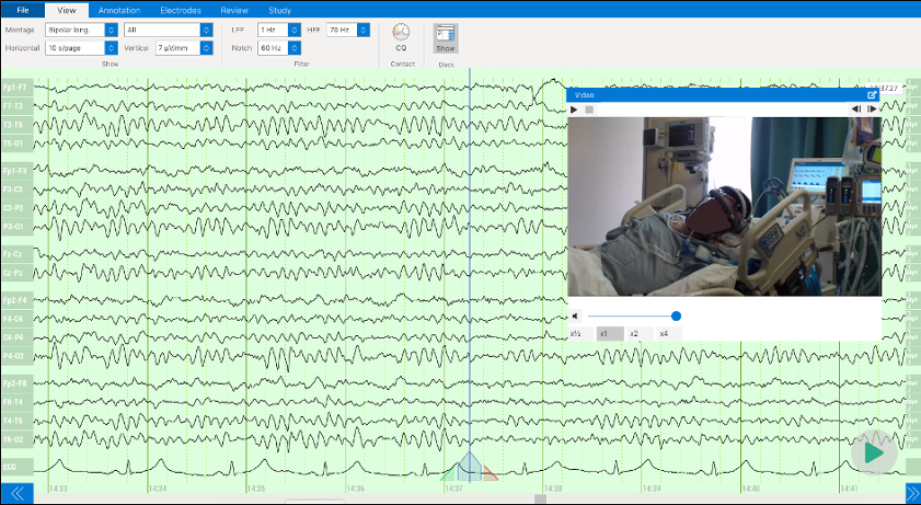
Video EEG is an important tool in diagnosing and monitoring patients with epilepsy or other neurological conditions. It helps distinguish physiologic or external artifacts from epileptic seizures or epileptiform discharges associated with seizures. Video EEG also helps to differentiate epileptic seizures from psychogenic nonepileptic spells that are mistaken for seizures and other episodic abnormal movements associated with other conditions, such as tic disorders, tremor, or periodic limb movements of sleep. Simultaneous video and EEG recordings enable the establishment of correlations between abnormal movements and abnormal wave activity.1,2,3,4
For awake patients, video can identify sources of noise such as muscle artifacts, blinking, chewing, forehead wrinkling, and even ear wiggling. For unconscious patients, video can identify external sources of artifact, such as electronic devices in the ICU (for example, an artificial respiration device). The inclusion of video in the EEG improves the ability to accurately diagnose a patient. Additionally, video can confirm correct electrode placement and headset positioning.
In this blog, we discuss the benefits of using video integration in EEG and what Zeto EEG can offer.
Different noise sources in EEG
EEG recordings can be impacted by various sources of noise and interference, including physiological noise, environmental noise, EEG electrode placement, electrical artifacts from EEG electrode malfunctions or from external electrical devices, and motion artifacts. These artifacts can make it difficult to distinguish genuine brain activity from noise, compromising the ability to accurately interpret EEG recordings.
Physiological noise arises from the body’s internal processes like muscle contractions, heartbeats, and eye movements, generating electrical signals that can interfere with the EEG recording. Environmental noise, on the other hand, encompasses external disturbances like electrical noise from equipment, electromagnetic interference, or ambient noise, which can introduce unwanted signals into the EEG data. Electrical artifacts can also be produced by the EEG equipment itself, resulting from issues like poor grounding, incorrect amplifier settings, or malfunctioning electrodes. Lastly, motion artifacts arise from bodily movements such as head motions, jaw opening and closing, or eye blinks, leading to distortions in the EEG signal and complicating accurate data interpretation.
To account for artifacts and enhance the interpretation of EEG recordings, video recording is often used in conjunction with EEG to identify and exclude sources of noise from the data.
Video EEG: Complement or Necessity?
Video is particularly useful for identifying artifacts related to movements or other physical activities that may produce electrical signals in the EEG recording. For example, if a patient moves their arm during an EEG recording, this movement can cause changes in the electrical activity picked up by the electrodes, which is recorded in the EEG. By observing the video recording, it is possible to identify the movement as an artifact and avoid misinterpreting the EEG data. This helps ensure that EEG signals identified as epileptiform activity represent genuine brain activity, rather than artifacts from other sources. Epileptiform waves can sometimes be difficult to distinguish from artifacts, as they can have similar characteristics in the EEG recording.
Video can also help diagnose and classify epileptic seizures and distinguish them from psychogenic nonepileptic spells and episodic abnormal movements due to other medical conditions that are not epileptic seizures.1,2,3,4,5 For example, if a patient experiences an epileptic seizure during an EEG recording, the video can help correlate the EEG signal with the visible movements and behaviors of the seizure. A psychogenic nonepileptic spell will show motion artifacts on the EEG but will not show underlying abnormal brain wave activity. Furthermore, certain behaviors such as squeezing the eyes shut while shaking, awareness during the seizure, and returning to baseline mental status right after the event can suggest a psychogenic cause.5
Sometimes tics, tremors, muscle twitching, and other abnormal movements in the tongue or a limb or digit can be mistaken for epileptic seizures, particularly in the ICU setting where subclinical seizures occur more frequently. Subclinical seizures are epileptic seizures that are not associated with characteristic abnormal movements usually associated with seizures. Video EEG can determine if the suspicious subtle abnormal movements are in fact due to epileptic seizures in most cases. If the abnormal movements represent subclinical epileptic seizures, they would be expected to correlate with epileptic seizure brain wave activity on the EEG.
Video recording provides additional information about the patient’s behavior or movements at the time of a seizure or seizure-like event, which is called a seizure’s semiology.1,3,5 Gaze deviation and arm stretching right before the seizure or at seizure onset can point to the seizure origin in the brain and inform localization of seizure foci relative to brain hemispheres or specific areas such as the motor cortex, supplementary motor areas, and frontal and parietal eye fields. Seizure semiology also includes the subjective sensations and experiences immediately preceding and during a seizure. It is critical to the diagnosis and classification of seizure disorders, which in turn affects the treatment plan. A video EEG is critical to presurgical evaluation prior to epilepsy surgery to diagnose and classify epileptic seizures when present and avoid unnecessary surgery due to psychogenic nonepileptic spells or other episodic abnormal movements not due to epileptic seizures.1
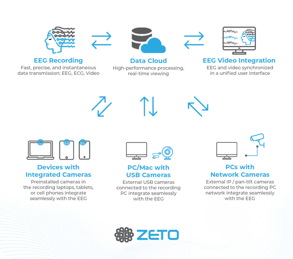
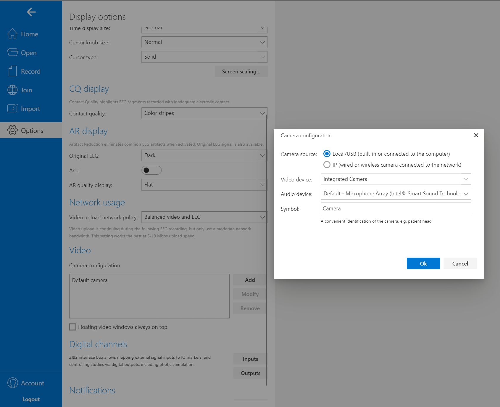
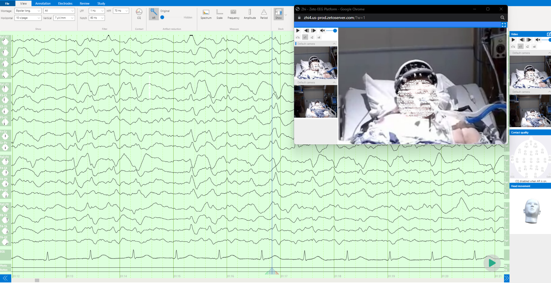
Zeto EEG: Seamless Compatibility with Any Camera, Reimbursable, Supports Up to Four Cameras
Zeto’s video integration feature reshapes the users expectations of what is possible with modern technology. The Zeto system enables simple camera controls (zooming and panning) through the cloud, ensuring simple usability.
One of the most appreciated features by users of the Zeto platform is its compatibility with any integrated or USB enabled camera. That eliminates the need for additional camera purchases and helps operators to use Zeto EEG more easily across a wide range of settings and recording locations. The Zeto Cloud Platform supports the use of up to four cameras simultaneously, providing medical professionals with the flexibility to monitor patients from multiple angles.
In addition to its compatibility with integrated and external USB cameras, Zeto offers the simple integration of IP cameras, whether they are wired or wireless. This capability is particularly beneficial when medical personnel already have pre-installed IP cameras in the room, as they can add these cameras to the Zeto Cloud Platform for a seamless integration. IP camera integration often depends on existing local IT infrastructure and cybersecurity guidelines which can result in longer integration timelines compared to integrated or USB – based camera solutions. But with the added configuration time come additional conveniences and features (such as lowlight or night vision capabilities) which often make this extended setup more than worth it.
Manually able to point or use of remote pan/tilt features
Automatic or manual zoom options available
Available in most devices
With any of these camera options, maximum image resolution depends on the utilized camera hardware, but integrated, USB, or IP camera options are available with Ultra 4k resolutions or higher. The consideration then becomes data storage size and related storage costs more than the technical ability – providers may still choose to save data in lower resolution settings simply to save storage costs.
Another powerful tool of the Zeto Cloud platform is the ability for the clinician to remotely access the recording through any device connected to the internet. Allowing for real time analysis when the provider is not present in the recording environment.
Medical practitioners will find the real-time monitoring capability invaluable, enabling them to observe patients with precision. Additionally, Zeto’s video EEGs, utilizing the integrated cameras, are eligible for reimbursement through CPT codes, specifically for EEGs lasting 24 hours or longer.
By utilizing video, Zeto ensures accurate positioning of the headset and proper electrode placement. This attention to detail guarantees the collection of reliable and precise EEG data.
In conclusion, video recording is an important complement to EEG recordings. It helps identify and exclude sources of artifact in EEG and confirms the presence of epileptic seizure activity and epileptiform discharges important for the diagnosis and classification of epileptic seizures. Video EEG also helps to identify psychogenic nonepileptic events and episodic abnormal movements from other medical conditions mistaken for epileptic seizures. By using video in conjunction with EEG, clinicians can improve the interpretation of EEG recordings, leading to more accurate diagnoses and better treatment outcomes for patients with neurological conditions.
Zeto’s video integration feature enhances convenience and accuracy, ultimately resulting in better diagnosis and treatment outcomes for patients.
Prestigious International Annual Awards Program Recognizes Standout Digital Health & Medical Technology Products and Companies
SANTA CLARA, Calif., – May 22, 2023 – Zeto, Inc , a commercial-stage medical technology company transforming EEG brain monitoring for health care, today announced that it is the recipient of the “ Best New Technology Solution for Neurology ” award in the 7th annual Awards program conducted by MedTech Breakthrough, an independent market intelligence organization that recognizes the top companies, technologies, and products in the global health and medical technology market.
Zeto makes it possible for any healthcare facility to offer a convenient, turnkey EEG solution to their patients. The company provides the first and only FDA-cleared, wireless, dry EEG headset that can be put on by any medical staff within five minutes.
The Zeto Cloud provides high-performance, real-time viewing, analysis, and reporting. Live video and data can be interpreted seamlessly via the cloud, and is further enabled by AI-based, FDA-cleared seizure detection. Zeto also offers optional continuous monitoring by certified EEG technologists, and interpretation services by board-certified neurologists.
EEG device innovation, important among current neurology solutions, highlights include active electrode technology, 10-20 positioning, soft, flexible electrodes, comfort for patients, advanced noise shielding and noise cancellation, and clinical-grade signal quality. Built to feel like a bike helmet, the device offers easy head size adjustment via adjustable dials.
“The massive unmet need for brain monitoring extends beyond neurology departments to ICUs, EDs, remote monitoring, telehealth, and physician offices. At Zeto, we want to eliminate any negative experience with a product that is both convenient for the operator and comfortable for the patient,” said Aswin Gunasekar, CEO of Zeto, Inc. “Receiving the ‘Best New Technology Solution for Neurology’ award from MedTech Breakthrough is a testament to the value our product is delivering.”
The mission of the MedTech Breakthrough Awards is to honor excellence and recognize the innovation, hard work, and success in a range of health and medical technology categories, including Telehealth, Clinical Administration, Patient Engagement, Electronic Health Records (EHR), Virtual Care, Medical Devices, Medical Data and many more. This year’s program attracted more than 4,000 nominations from over 17 different countries throughout the world.
“The portable nature of this EEG headset from Zeto has amazing potential for patients and practitioners, especially as it offers similar signal quality to traditional devices,” said James Johnson, managing director, MedTech Breakthrough.
“EEG is an important diagnostic technique for several neurological issues. However, the biggest limiting factor is that conventional EEG needs a trained operator to administer the test, and if a facility doesn’t have a neurologist on call, they are unable to read the EEGs. Congratulations to Zeto for transforming the way we perform EEGs, with tangible and real results.”
Zeto, Inc. is an award-winning, privately held medical technology company located in Santa Clara, CA, that is focused on transforming the way electroencephalography (EEG) is performed at hospitals and clinics. Zeto’s revolutionary FDA-cleared EEG headset and cloud platform bring the traditional EEG procedure to the 21st century. The company plans to leverage its hardware and software technology to improve noninvasive monitoring of the brain’s electrical activity and achieve better outcomes for neurological conditions such as epilepsy, sleep disorders, autism, stroke, and concussion.
To learn more about Zeto’s neurology solutions including remote EEG monitoring, please visit: https://zeto-inc.com or email us at info@zetoinc.com.
Event Related Potentials (ERPs) in EEG provide insight into how our brain processes information, reacts to its environment and adapts to challenges. ERPs differ from the traditional clinical tradition of evaluating continuous spontaneous brainwaves in patients. With ERPs, experimenters can examine the brain’s response to succinct events. For a summary on ERPs, see here.
One of the most crucial aspects in capturing clean ERPs is knowing precisely when specific target events occur. So-called event markers are commonly used to timestamp the onset of target events in the continuous EEG tracings to enable further data processing. The target events can have different modalities and can either be initiated or perceived from the person receiving the EEG.
For example, participant initiated movements are known to elicit robust ERPs.12 However, more commonly, ERPs are recorded from participants perceiving sounds, language, images, or smells.3,4,5In principle, ERPs will emerge via subsequent processing as long as experimenters established a reliable method to repeatedly mark the onset of such events in the EEG.
There are two technical aspects that determine the quality of ERP event markers:
Delay: Time from when an event occurred to when it is marked in the EEG data
Jitter: Consistency of the delay with which the event is marked in the EEG data
Long delays with large jitter complicate EEG analysis up to the point in which the targeted ERP component becomes unobtainable or requires too many trial repetitions to appear. Short delays with minimal jitter create the ideal technical conditions to obtain ERPs with a minimally possible amount of trial repetitions.
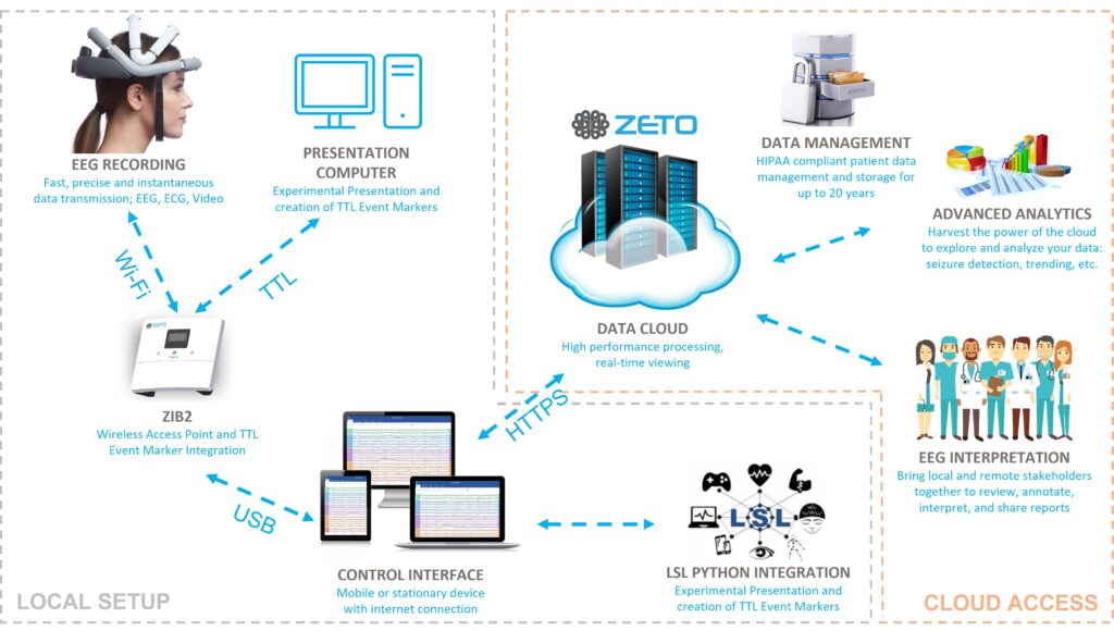
With Zeto’s event marker integration, users benefit from accurate event marker timing and distributed cloud data access and management – see Figure 1.
Figure 1. Schematic diagram of Zeto’s local event marker integration and remote data streaming and management features. Event markers from the presentation computer timing are merged with the EEG data locally via the Zeto Interface Box 2 (ZIB2) and then passed on to the Zeto Cloud. A simultaneous LSL integration and related multi-model data recording become possible out of the box while maintaining Zeto’s existing cloud streaming, data, and user management features.
ZETO ERP FEATURES
The Zeto EEG platform offers the ability to integrate external markers wirelessly at an 8-bit resolution within a 2 ms delay and less than 1 ms technical jitter. In other words, the user can distinguish between 255 unique event markers that they can repeat as closely as 4 ms from one another. With these features, Zeto EEG is equipped to reveal accurate auditory, visual, and senso-motoric ERPs across a wide range of applications.
Event markers are collected by the system via an 8-bit DB9 connector built into the Zeto Interface Box Version 2 (ZIB2) using Transistor-Transistor-Logic (TTL) signals.6 The ZIB2 acts as a data access point for the wireless Zeto WR19 headset. Synchronization between the ZIB2 and Zeto WR19 is handled at a nano-second range, eliminating both the delay and jitter introduced by the wireless data transmission. Incoming event markers are retroactively aligned with the data point at the time of collection.
Users can extract the event marker data from the Zeto system in multiple ways:
1) EDF+ File Zeto users can export finished EEG recordings in various ways but the most popular is the EDF+ file format that is readable by most common EEG analysis tools. Event markers appear in the EDF+ file as digital I/O channels synchronized with the EEG data. Some EDF readers will also display the embedded event markers in the viewer.
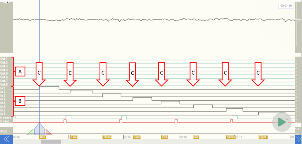
2) Visualization In the Zeto cloud software, users can visualize the TTL event marker inputs along with the EEG by selecting the “ALL” montage in the montage menu. This view is particularly useful for troubleshooting when setting up the ERP experiment. Offline or in real-time the user will see incoming event marker codes visualized high or low values in separate channels. The event marker channels are simultaneously translated into event marker labels that co-appear at the bottom of the screen (Figure 2).
Figure 2. Close-Up of the “All” Display montage: Output (“A”) or Input (“B”) event marker channels for 8 bits each, translated into up to 8-bit (255) unique event marker labels. The event marker mapping can be freely configured and labeled as desired prior to the recording. In this example, eight input event marker signals are embedded in the data file (pins 1 to 8) and show up as square waves (“C”). Each input event marker channel is mapped to an annotation, labeled “One” through “Eight” respectively at the bottom of the data screen.
3) Real-time lab streaming layer (LSL) Export Eight digital input and output channels each are made available via lab streaming layer (LSL) API in real-time, enabling the user to note the stimulus onset directly in the data stream. Event makers and EEG are synchronized and merged prior to providing this data to the LSL streaming socket. As a result, event timing and EEG data remain perfectly synchronized even if there are LSL related streaming delays. Additional LSL API synchronization features remain available to users for additional real-time data integration.
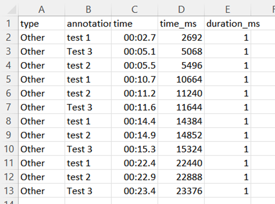
4) Offline Event Marker Files Users have the option to export marker files after the recording is completed. That marker file contains precise marker timing and label information for all event markers recorded for easy processing in third party analysis tools such as MATLAB, ERPLAB, Python or others. This allows for separate analysis of event data and EEG data found in the exported EDF+ file.
The event file can be exported in “.csv” format (Figure 3), or a comma-delimited format called “.zmrk”. Both are compatible with most common EEG processing tools currently available for research.
Figure 3. Event Markers listed in .csv format.
ZETO EVENT MARKER TIMING VALIDATION
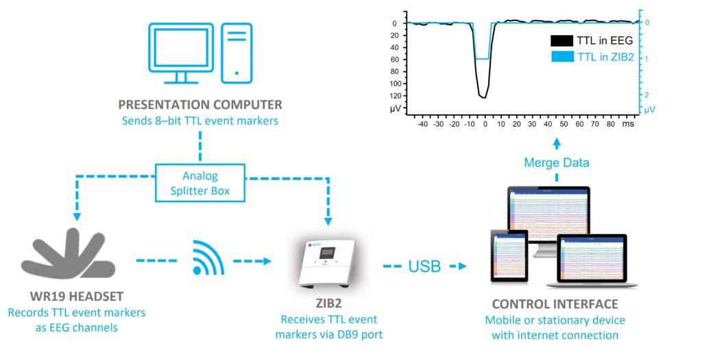
Zeto validated the event marker timing to establish the delay and jitter attributes under working conditions. To do this, a testing setup split the incoming TTL trigger voltages via an analog splitter into two exact 8-channel copies. One copy of the event marker signals got connected to the ZIB2 input trigger ports while the second copy got connected to 8 channels of the WR19 headset. As a result, incoming event markers both appeared as digital events in the datastream and voltage changes in the EEG channels (Figure 4). Subsequent processing revealed the real-life delay and jitter between the incoming event marker signals and the EEG recording.
Figure 4. Schematic of the event marker timing test setup. The presentation computer sends 8-bit TTL event markers to an analog splitter box. One copy of the TTL signals arrives at the ZIB2 and gets converted into event labels. The other copy arrives at the headset and feeds into 8 of the EEG channels to show up as signals in the EEG data file.
Using this approach, event marker timing was established as stable, at < 2 ms delay and <1 ms jitter, which is a good basis to reliably capture ERP signals in EEG.
ZETO’S STIMULATION AND SYNCHRONIZATION PLATFORM PARTNERS
It is important to note that the Zeto system provides extremely precise synchronization on the receiving end of the event marker only. In fact, a much more likely source of both delay and jitter in ERP experiments occurs during stimulus presentation and subsequent event marker generation.
To avoid timing complications that occur prior to event markers entering the Zeto system, Zeto has partnered with two stimulus and synchronization platforms – both tested with our products. These stimulus presentation and synchronization products are designed to eliminate delay and jitter on the event marker onset. In addition, they offer a variety of additional functions, including experiment writing and presentation software, participant response boxes, and photodiodes.
Both partners have implemented out-of-the-box integrations for Zeto and are ready to service Zeto customers.
Cedrus devices are designed for precise, jitter-free event marking and fit a variety of budgets. SuperLab is an experiment writing application, while software support for their hardware interfaces also includes Matlab, E-Prime, Python, C++, etc.
Psychology Software Tools isa prominent software and hardware company that helps researchers address challenges in human behavioral studies. PST are creators of E-Prime, a market-leading experiment writing platform.
Sean McWeeny, Elizabeth S. Norton. “Understanding Event-Related Potentials (ERPs) in Clinical and Basic Language and Communication Disorders Research: A Tutorial.” PMC. https://www.ncbi.nlm.nih.gov/pmc/articles/PMC3016705/
Zeto, Inc., an innovative EEG brain monitoring company, announced its participation in the upcoming Vision Sciences Society (VSS) annual meeting in St. Pete Beach, Florida, from May 19th to 24th.
The Vision Sciences Society (VSS) Annual Meeting is a premier gathering that covers the broad scope of vision science. The meeting brings together experts from various disciplines, including visual psychophysics, visual neuroscience, computational vision, and visual cognition. As part of this event, Zeto, Inc. will showcase its latest product features tailored for EEG in clinical research applications.
Originally developed for the rapid acquisition of clinical EEGs in hospitals and clinical practices, Zeto is responding to an increasing number of requests from researchers and clinical research organizations (CROs) who share a common need for high-quality EEGs recorded more efficiently compared to traditional technologies.
The research features introduced by Zeto include the ability to record timing-precise event- related potentials (ERPs) through their unified cloud platform. Users can now benefit from the ease and security of Zeto’s cloud interface yet maintaining sub-millisecond precision on input and output event markers which are crucial for high-end ERP data acquisition. This cloud platform is particularly relevant for CROs and large-scale multi-site research projects, providing a central place for data access and management.
With advanced API features, state-of-the-art encryption and user management, researchers can conveniently interact with EEG and synchronized video data using commonly available scripting languages such as Python®, MATLAB®, or most other programming languages. This significantly speeds up the ability to process large multi-site datasets and fosters collaboration and synergies among multi-center stakeholders.
“Zeto has been providing tremendous value for our clinical customers in hospitals and private practices, but we are particularly excited to now offer the convenience of Zeto EEG to CROs and cutting-edge researchers,” says Florian Strelzyk, Chief Sales Officer at Zeto. “The ease of use, comfort, and refined feel of our products will open up a wide range of new opportunities to bring EEG out of the lab and into research environments that are closely related to the topics they study. Imagine the possibilities ahead with a platform that is quick to apply and easy to work with. Successful research frequently relies on large teams extracting clinical findings efficiently and Zeto can provide this multi-user experience like no other tool currently available.”
About Zeto
Zeto, Inc., is an award-winning, privately held medical technology company located in Santa Clara, CA focused on transforming the way electroencephalography is done in clinical and research settings. Zeto’s revolutionary FDA-cleared EEG platform brings the traditional EEG procedure to the 21st century by offering the WR19, a zero-prep, wireless, easy-to-wear headset with active, dry electrodes that can be positioned as per the 10-20 system. The Zeto headset is backed by a cloud data and software platform, a real-time LSL-based API, and a TTL-based trigger device for ERP studies. The company plans to leverage its platform technology to improve access and quality to medical EEG testing and to enable and improve adjacent biomedical research and clinical trials.
As a healthcare professional, you know how important it is to have reliable equipment that can withstand the daily grind of a busy medical practice. That’s why we designed Zeto EEG – a rugged, clinical-grade headset that is built to last.
Zeto’s durable EEG headset is made from high-quality, clinical-grade materials that are easy to clean and maintain. It has a light but substantial feel, making it comfortable to wear for extended periods of time. But don’t let its light weight fool you – Zeto’s resistant EEG headset is tough enough to handle collecting EEG data in even the most demanding clinical environments.
To back up our commitment to quality, we offer up to 4 years hardware warranty that covers intended use. Customers have the choice to purchase this warranty at once upfront or extend it annually. This means that you can use Zeto with confidence, knowing that it is built to last. Excluded from that warranty is unintended use such as submerging or washing, sitting on, tearing, or intentionally over-bending the headset.
Our standard Service Level Agreements (SLAs) provide 72 hours replacement assurance. For those who need even faster replacement, our premium SLAs assure that a replacement headset can be with you within 24 hours during weekdays as long as we receive your request by 2 p.m. (ET).
Curious about how well our Zeto’s reliable EEG headset can withstand the daily wear and tear of a clinical setting? We put our product to the test with a drop test, simulating the accidental drops and impacts that can occur during everyday use.
Watch the video to see just how rugged and durable Zeto truly is:
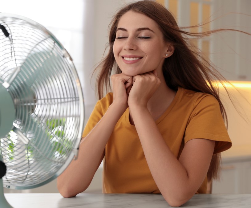
Sweat artifacts are a common problem in electroencephalography (EEG) recordings. They can noticeably affect the quality of the recorded tracings and make it difficult to read the underlying EEG signals. Sweat artifacts in EEGs occur when the body’s biological sweat response alters the conductivity of the skin in a way that affects the electric signals picked up by the electrodes. Such changes not only occur when sweat is visible on the scalp but also occur when the body heats up and prepares to sweat.
In this blog, we will explore the causes of EEG sweat artifacts, their effects on EEG recordings, and strategies for mitigating their impact.
What Causes Sweating
Sweat is crucial for human thermoregulation and can be caused by a variety of factors, including anxiety, nervousness, or physical exertion. Biological changes during menopause also increase the chance of sweating. Regardless of these and other factors, a warm testing environment is the main driver of sweating.1 Systematic studies revealed that temperatures above 79°F (~26°C) can have a noticeable effect on the EEG and signal morphology.2
The Biology of Sweat Artifacts in EEG
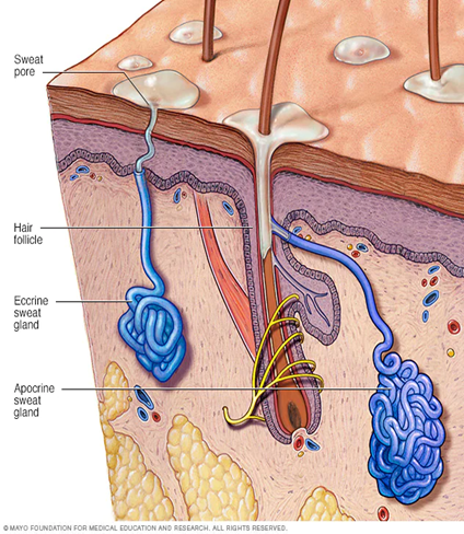
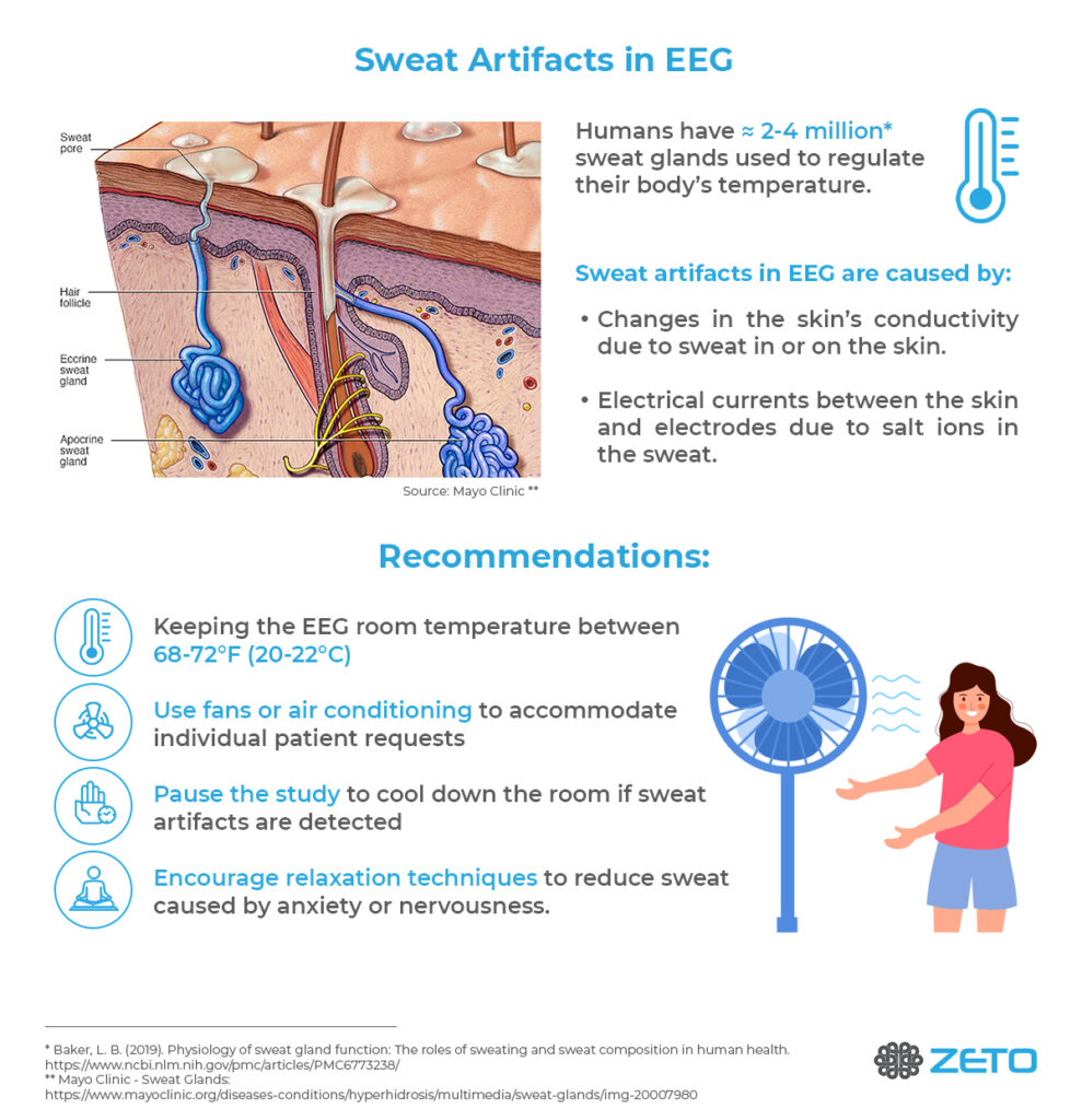
Sweating is not simply the appearance of sweat on the skin but the result of a cascade of biological changes that lead to the skin’s ability to secrete liquid from the sweat glands, onto the skin (Figure 1).
The filling of the sweat glands with liquid in preparation of sweat excretion increases the electrical conductivity of the skin rapidly which affects the morphology of the EEG signals. These rapid changes in skin conductivity and the uneven distribution of the sweat glands across the skin result in recordings prone to major EEG artifacts, with single channels showing large signal changes at different times and locations.3
Figure 1. Cross section of epidermis and dermis skin layers with embedded hair follicle, eccrine, and apocrine sweat glands. Source: Mayo Clinic
The Physics of Sweat Potentials in EEG
In addition to biological changes in the skin’s conductivity, the composition of the sweat itself is contributing to electrical potentials that EEG amplifiers pick up. Sweat contains high sodium chloride and lactic acid which react with metallic components of the EEG electrodes, generating electrical potentials.4 These electrical potentials combine with skin and sweat gland potentials into what is visible in the EEG as sweat artifacts.
Appearance of Sweat Artifacts in EEG
Sweat artifacts in EEG can appear in various morphologies or shapes that are affected by biological factors such as the severity and generality of the sweat response. The sudden onset of sweating across the entire body will appear different from sweating that occurs over time or may be more limited by body part or region. More relevant for the appearance in EEG though, are the analog or digital filter settings of the recording.
Amplifiers with a built-in low-frequency hardware filter will show a more subdued sweat artifact even without any digital filtering. True direct current (DC) amplifiers that do not have any analog low-cut-off filter will show the build-up to a sweat artifact in their raw data much more because small changes over time can be picked up much better.
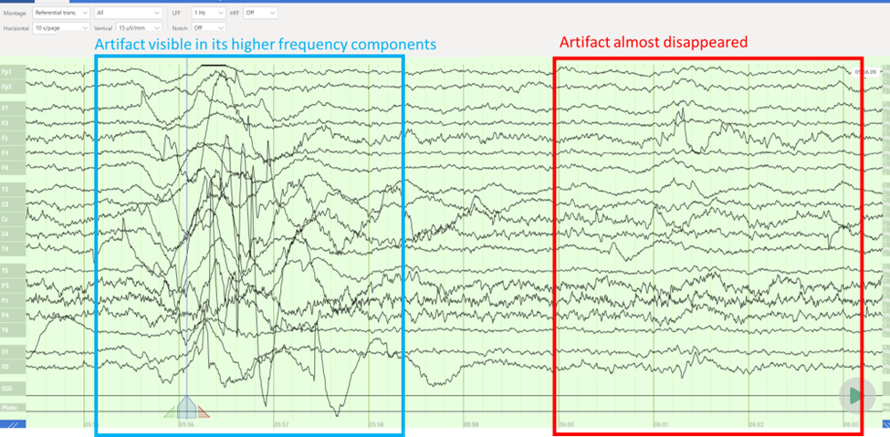
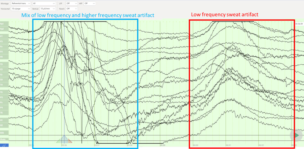
Most clinical EEGs are viewed at a 1 Hz–70Hz bandpass filter as recommended by ACNS.5 EEG Sweat artifacts viewed using a 1 Hz low-cut-off filter generally show up as slow wave components around a 1 Hz–3 Hz frequency in otherwise normal background activity; for an example, see Figure 2. Disabling the low-cut-off filters, however, exposes additional low-frequency drifts related to sweat that are otherwise masked by digital signal processing; for an example, see Figure 3.
Figure 2. Filtered sweat artifact in a full 19-channel clinical EEG viewed in a referential montage. 1 Hz low-frequency forward Butterworth filter applied. The slow meandering signal drifts almost completely disappears after filtering (red frame).
Sharper signal drifts remain visible even after filtering (blue frame). For most clinical recordings, EEG tracings such as this are indicators of the biological changes that are caused by a sweat response. Data was recorded using Zeto’s WR19 headset at 79°F (~26°C).
Figure 3. Unfiltered sweat artifact during the same data segment, as presented in Figure 2. Slow meandering (red frame) and at times sharper signal drifts (blue frame) reflect the biological changes in the skin’s conductivity due to sweating.
How to Get Rid of Sweat Artifacts in EEG
There are two common ways to reduce or avoid sweat artifacts in EEG recordings.
EEG operators can reduce the biologically triggered changes that lead to sweating. In preparation for the EEG recording, operators can ask patients to avoid strenuous exercise, caffeine, and alcohol prior to a scheduled EEG session, ideally 24 hours before the test. During EEG recordings, Kappenman and Luck recommend maintaining a cool temperature in the recording environment to minimize the occurrence of EEG sweat artifacts. They recommend a comfortable temperature of 68°F –72°F (20°C –22°C) and using fans or air conditioning to prevent humidity buildup.2
During the EEG session, EEG operators should assure the best possible electrode contact with the scalp to reduce skin impedance under the electrode. In traditional amplifier systems with wired electrodes, this can be achieved via additional skin preparation and abrasion. With active quick-apply EEG recording systems, such as Zeto’s headset, operators can assure proper electrode landing with each conductive leg touching the scalp.
Bottom Line – Recommendations
In hectic clinical day-to-day EEG schedules, the easiest way to avoid sweat artifacts in most patients is to avoid sweating in the first place. For that reason, option #1, mentioned previously (reducing sweating), is the most robust way to assure consistent EEG data quality.
Keep the EEG room temperature at 68°F – 72°F (20°C–22°C), especially when recording unconscious patients who cannot communicate their comfort levels; keeping an optimal temperature reduces the body’s need for sweating.
Use fans or air conditioning to accommodate individual patient’s temperature requests. Each patient is different; ask to make sure they are not hot.
If EEG sweat artifacts are detected, consider pausing the study to cool down the room (i.e., opening the door, reducing the room temperature, and/or the use of a fan).
Relaxation techniques: Encouraging the patient to relax and breathe deeply. This can help to reduce sweat caused by anxiety or nervousness.
By implementing these strategies, EEG operators can help minimize sweat artifacts in EEGs and obtain cleaner results. It is important to work closely with the patient and monitor the EEG tracings for any signs of EEG sweat artifacts during the test to more adequately address issues with data quality.
Kalevo, L., Miettinen, T., Leino, A., Kainulainen, S., Korkalainen, H., Myllymaa, K., … & Myllymaa, S. (2020). Effect of sweating on electrode-skin contact impedances and artifacts in EEG recordings with various screen-printed Ag/Agcl electrodes. https://ieeexplore.ieee.org/stamp/stamp.jsp?arnumber=9017959
Siddiqui, F., Osuna, E., Walters, A., Chokroverty, S. (2006). Sweat artifact and respiratory artifact occurring simultaneously in polysomnogram. https://pubmed.ncbi.nlm.nih.gov/16461004/
Greenwood Leflore Hospital in Greenwood, MS has recently upgraded its EEG capabilities with the implementation of a modern solution. The hospital, which has always prioritized offering in-house EEG tests for its patients, has adopted a technology that allows for a faster and more efficient EEG experience.
Patients can now benefit from a shorter setup time and a more convenient testing process, without the need for skin prep or gel. The new technology has received positive feedback from patients, who appreciate the faster scheduling, the lack of need for post-test hair washing and simplified testing process.
“The new EEG solution has been a valuable addition to our hospital,” says Steven Robinson, Director of Cardiopulmonary Care at Greenwood Leflore Hospital. “It offers a more efficient and patient-friendly approach to EEGs, while also providing high-quality results.”
“We are proud to support the improvements to patient care at Greenwood Leflore Hospital through the implementation of our EEG solution,” says Florian Strelzyk, Chief Sales Officer at Zeto, Inc. “Providing on-site testing is a crucial aspect of quality patient care and we are pleased to be able to contribute to this.”
The hospital’s ability to perform routine and urgent EEGs at a moment’s notice has also been improved, allowing for better patient care and a more streamlined experience. The EEG headset simplifies the testing process for both patients and healthcare providers.
In conclusion, Greenwood Leflore Hospital’s upgrade to its EEG capabilities has made EEG testing a quick and comfortable experience for patients, further enhancing the hospital’s commitment to providing top-notch care.