Zeto's wireless EEG machine

Wearable EEG Headband

Zeto to Showcase Technology to Make EEG Brain Monitoring More Accessible for New Providers and in Rural and Underserved Communities at 2022 AAN Annual Meeting

Today, Zeto, Inc. announced its participation in the 2022 American Academy of Neurology Annual Meeting, an event for neurologists and neuroscientists, taking place April 2-7, 2022.

The AAN Annual Meeting strives to promote the highest quality patient-centered neurologic care, assert the value of neurology to policymakers and other major stakeholders, and expand the neurology workforce to meet future needs for patient care.

“We are excited to be back in person for the 2022 AAN Annual Meeting. The past two years put a tremendous burden on many private practices and hospitals. In large parts of the U.S., hiring technologists required to record EEGs has been an everyday challenge due to the pandemic. That makes new providers or providers in rural and traditionally underserved communities struggle to offer crucial EEG tests at their own locations.

“For that very reason, Zeto will showcase our FDA-cleared EEG monitoring technology that enables providers to offer brain monitoring even without traditionally trained EEG technologists. Zeto EEG technology helps save valuable time and resources, while not compromising clinical standards. Neurologists can perform an EEG rapidly, even without an EEG technologist on site. At no upfront costs, and for as low as $65 per patient, providers can record full-montage EEG in under five minutes at the same quality as conventional gel EEG. Included in that per-patient fee are an EEG amplifier, consumables, cloud software with storage, video integration, warranty, training, and a powerful report writing tool. Optional EEG reading services further support clinical decision-making. In the past two years, we saw countless examples of how Zeto technology helped support providers to make EEG more available even under the most challenging circumstances,” said Florian Strelzyk, Chief Sales Officer at Zeto.

According to statistics, one in three patients recovering from Covid-19 may experience neurological or psychological effects from their infections, reflecting a growing consensus that the disease can have lasting effects on the brain.

The AAN Annual Meeting is the largest meeting and exhibition in the world to share common scientific and clinical interests in neurology.

About Zeto

Zeto, Inc. is an award-winning privately held medical technology company located in Santa Clara, CA, focused on transforming the way electroencephalography (EEG) is performed at hospitals and clinics. Zeto’s revolutionary FDA-cleared EEG headset and cloud platform bring the traditional EEG procedure to the 21st century. The company plans to leverage its hardware and software technology to improve noninvasive monitoring of the brain’s electrical activity and achieve better outcomes for neurological conditions such as epilepsy, sleep disorders, autism, stroke and concussion.

To learn more about our products, please visit: www.zetoinc.com or email us at info@zetoinc.com


Source: https://www.newswire.com/news/zeto-to-showcase-technology-to-make-eeg-brain-monitoring-more-21675785

The Role of EEG for Epilepsy Diagnosis, Management and Classification

People with epilepsy typically experience recurrent seizures. Despite the diverse causes of seizures, the common mechanism linking many types of epilepsy is the disruption of the brain’s normal electrical activity, which temporarily halts communication between neurons.

About 60% of epilepsy cases have a cause, a lesion, or abnormality in the brain, detectable by neuroimaging methods [1,2]. Another class of pathogenesis of numerous epileptic symptoms is an abnormal expression of specific receptors in the brain, which leads to increased excitation and decreased inhibition resulting in enhanced neural activity.

Because epilepsy can only be diagnosed based on electrophysiological evidence (detection of two independent epileptic events by EEG tests) the use of EEG is mandatory for epilepsy diagnosis and management. Furthermore, based on the EEG evidence a trained epileptologist can determine the type of seizure and diagnose the type of epilepsy syndrome of the patients. The exact diagnosis can help to provide effective antiepileptic medication and prognosis.

Let’s find out the role of EEG in diagnosis, classification, and management in more detail. But first, let’s discuss what an EEG is.

What is EEG?

Electroencephalogram (EEG) is non-invasive research and diagnostic tool used to measure the changes of the brain’s electric potential over time, commonly called brain waves. This electric potential is generated by the discharges of millions of neurons. Although EEG does not have the spatial resolution of detecting the discharges of individual neurons, it can discern levels of activity associated with the major lobes of the human cerebral cortex. In other words, the EEG is a test that helps to detect electrical activity and abnormalities in a patients’ brain and localize them with a certain precision sufficient to make a diagnosis. An EEG equipment uses small sensors (electrodes) made of a conductive material attached to the scalp or they contact the skin. Often these electrodes are preconfigured inside an EEG headset to speed up the positioning.

Typically, specialists, clinical neurophysiologists, neurologists, and researchers carry out an EEG recording. Traditionally it has been done in clinics or academic laboratories and also has been adapted for home monitoring. While EEG has numerous research applications from basic research to Brain-Computer-Interface (BCI), in the field of clinical neurology it is mainly used to diagnose and monitor epilepsy and sleep disorders.

Diagnosis, Classification, and Patient Management

Diagnosis and treatment of epilepsy are often challenging. However, modern therapy provides many patients with multiple treatment options and often complete control of the seizure. After the first two seizures, evaluation should concentrate on:
1. Ruling out any non-epileptic medical or neurological condition that may generate seizures (e.g., psychogenic seizures)
2. Determine the type and location of seizures (e.g., focal, generalized, convulsive, non-convulsive)
3. Evaluating the relative risk of a seizure episode
4. Evaluating treatment options (e.g., diet, pharmacological treatment, surgical intervention, implanted control device)

The Use of EEG in Diagnosis of Epilepsy

Regardless of technological advancements, the first seizure episode typically is not captured in EEG. Numerous paroxysmal events can be confused with epileptic seizures, such as movement disorders, syncope, psychogenic seizures, etc. Probably, the most common event confused with epileptic seizures is syncope. To rule out non-epileptic seizures one needs to record abnormal activity from the brain as primary evidence. This is done by EEG equipment because all other methods to record brain activity are more expensive. At the same time, it is generally recommended to carry out a brain imaging study, such as magnetic resonance imaging (MRI). The MRI can reveal underlying cerebral lesions such as a tumor, stroke, vascular malformation, that could explain the seizure and also help localize it. However, not all epileptic seizures are associated with morphological differences in the brain that can be resolved by MRI. The class of epilepsy associated with electrographic seizures without visually observed MRI evidence is called non-lesional epilepsy.

The Vital Role of EEG in Epilepsy Diagnosis

Why does EEG play a central role in epilepsy diagnosis and treatment? Because EEG can:

● detect epileptiform activity,
● strengthen the putative diagnosis,
● identify the focal cerebral abnormalities, which may indicate a focal structural anomaly such as brain tumor, hemorrhage, vascular malformation and
● document particular epileptiform activity patterns linked to specific epilepsy syndromes

Trained clinicians can recognize a particular type of epilepsy based on their signature waveforms and distribution using an EEG device. Each type of epilepsy diagnosis entails specific treatment strategies. Typical EEG results provide a multiaxial diagnosis of epilepsy describing whether the seizure disorder is generalized or focal, symptomatic or idiopathic (unknown cause), or part of a particular epilepsy syndrome. Because no two epilepsy cases are identical, providing a detailed description of the type of epileptic waveforms, the topography (location in the brain), the frequency of occurrences, the triggering stimulus if there is any, and the effect of seizure on the cognitive and motor functions are all important aspects shaping the treatment strategy.

One critical aspect of epileptic seizures that can be captured by an EEG study is whether it is generalized or focal. The two require completely different medication and treatment strategies. In the case of generalized seizures, abnormal synchronized discharges quickly spread to both cerebral hemispheres, while in focal seizures the abnormal discharges remain localized to a certain area or areas. To capture these events, one needs to spend hours or days with a continuously recording EEG because these events are rare unless it is triggered by a known stimulus (light, sound, touch, anxiety, hyperventilation, etc.).

Because of the scarcity and unpredictable nature of epileptic seizures, these events may not be captured in the clinic during the EEG. However, the description of a seizure by a witness combined with the patient’s self-report can complement the information available from EEG. Abnormal EEG activity patterns that indicate the potential for seizures are called inter-ictal events (sharp waves and spike and waves). These events play an important role in localizing and seizures. Today, a lot of attention is paid to interictal events as potential biomarkers of an impending seizure. One of the biggest machine learning challenges in medicine is to predict seizures based on the types and occurrences of these interictal events.

The role of EEG in Classification of Epilepsy

The classification of epilepsy and the recognition of diagnostic categories based on EEG is an ongoing, evolving process. The categories we use today are not the same as the ones we used 30 years ago, and they change as we understand the disease better. We tend to overclassify epilepsy syndromes as each is associated with particular EEG features. Therefore, it is the task of an internationally elected committee of experts ”International League Against Epilepsy (ILAE” to update the classification systems from time to time, based on consensus and published empirical evidence [3]. Because the classification is evidence-based, and evidence is subject to technological advances, the EEG and other methods, such as neuroimaging, molecular biology, and genomics have a great impact on the classification progress. And will be informed as times go on by developments in imaging, molecular biology, and genetics.

The role of EEG in Management of Epilepsy

The main objective for treating epilepsy patients is to control seizures entirely without causing undesirable side effects. Therefore, besides EEG being an indispensable part of diagnosis, it is also necessary for epilepsy management. Until today the primary measure of the efficacy of epileptic drugs was the extent it reduces seizure frequency. This assessment was often based on self-reports, diary, and caretakers’ notes. With the widespread availability of EEG, this is expected to change and EEG could be utilized for quantifying the efficacy of any treatment, from drug therapy to special diets.

Conclusions

Patients diagnosed with epilepsy have more therapeutic options available to them today than yesterday. To maximize the benefit of these options, clinicians must make an accurate diagnosis of epilepsy syndrome, select and use medications effectively, and promptly refer patients where necessary.

Among the broad range of available diagnostic methods, EEG is still the most versatile and affordable research and diagnostic tool that helps study the brain’s electrical activity and recognize patterns associated with epilepsy. Most importantly EEG provides detailed information about the type and localization of epilepsy.

While it has a very limited spatial resolution and is prone to misinterpretation, EEG remains the gold standard of epilepsy diagnosis. It is and it will remain in the equation to provide better care for patients and to feed our curiosity about the inner workings and communications of brain tissue.

References:

1. Nguyen DK, Mbacfou MT, Nguyen DB, Lassonde M. Prevalence of nonlesional focal epilepsy in an adult epilepsy clinic. Can J Neurol Sci. 2013 Mar;40(2):198-202. doi: 10.1017/s0317167100013731. PMID: 23419568.
2. Téllez-Zenteno JF, Hernández Ronquillo L, Moien-Afshari F, Wiebe S. Surgical outcomes in lesional and non-lesional epilepsy: a systematic review and meta-analysis. Epilepsy Res. 2010 May;89(2-3):310-8. doi: 10.1016/j.eplepsyres.2010.02.007. Epub 2010 Mar 15. PMID: 20227852.
3. https://www.ilae.org/guidelines/definition-and-classification/proposed-classification-and-definition-of-epilepsy-syndromes

A Guide to EEG Basics (Electroencephalography) & Devices Used

Every machine requires some circuitry or motherboard that controls the machine’s functions and operations. Likewise, humans also possess a complex computing system inside the body: the brain. The brain’s inner workings and connections are mysterious. An intricate system of neurons link together to form the brain’s jelly-like morphology.

Advancements in medical science and inventions have improved our understanding of how the brain works. One such invention was electroencephalography, a method and device used to record and analyze the electrical activity occurring inside the brain.

While the first EEG was performed in 1924, the technology is constantly evolving. Today, modern portable EEG devices are changing the way we look at the brain.

In this EEG guide, you’ll learn what an EEG machine is, what an electroencephalography is, how the EEG system operates, and what the various devices are used for.

What is Electroencephalography (EEG)?

Electroencephalography 1, or EEG, is a procedure used to measure and record the electrical activity of the brain in the form of waves. One can monitor the neurophysiological function of the brain while the subject is performing different tasks. Various electrical abnormalities can also be detected with precision.

As we understand the brain better, our EEG technology and the way we interpret the signals of the brain continue to improve. This has led to new ways of performing EEGs, such as wireless EEG systems that allow us to continue to learn the secrets of the brain.

What is an EEG?

Our brain is composed of billions of interconnected neurons. These neurons work by generating electrical potentials in the form of neuronal impulses which travel through the brain. EEG works on the principle of measuring these electrical potentials/voltages generated inside the brain.

What is an EEG Machine?

An EEG machine measures these electrical potentials by recording the differences in voltage between various points using a pair of electrodes. Then, the recorded data is sent to an amplifier.

The amplified data is eventually digitized and displayed on the monitor of the EEG machine as a sequence of voltage values that fluctuate in time. The resulting EEG waveforms from the EEG machine are interpreted to detect signs of abnormality inside the brain.

Parts of an EEG Machine

Essentially, an EEG machine is made up of the following primary device(s):

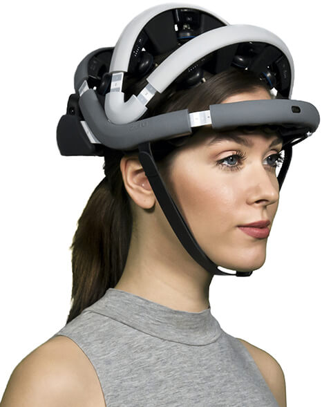
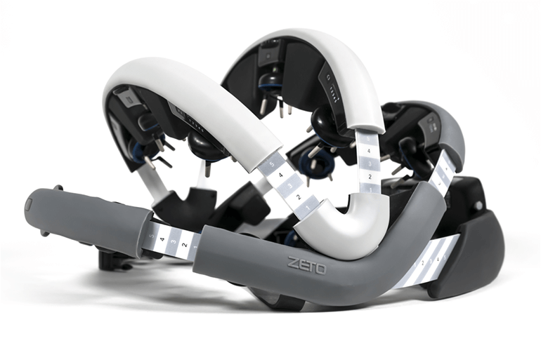
  • Electrodes: The electrodes pick up small electrical brainwaves produced by neurons. These are attached to the scalp with a special paste. Modern EEG machines possess a wearable cap with electrodes pre-installed inside the cap.
  • Amplifiers: As the signals travel from the electrodes through the machine, they run through an amplifier that boosts the incoming signal enough to be displayed on the screen.
  • Computer Control Module: The amplified signals are processed by a computer.
  • Display Device: The processed signals are displayed on the screen to be analyzed by the operator. Before the digital monitoring methods became prevalent, waveforms were plotted with a moving pen on rolls of graph paper.3

How is an EEG performed?

An EEG test may be performed either as an outpatient study or as part of your stay in the hospital. Various EEG technology and techniques are used depending on your health condition. Generally, an EEG procedure utilizing EEG technology is done in the following way:

  • The patient is asked to relax by lying on a bed or sitting in a chair.
  • Various electrodes (between 16, 20, or more) are attached to the scalp using a special electrolyte paste, or the patient is fitted with a cap containing the electrodes.
  • The patient is then asked to close their eyes and remain still.
  • Generally, an EEG technologist performs this procedure, which may take from 20 minutes to 2 hours, not including the electrode prepping.
  • Longer brain monitoring requires the patient to be admitted to the hospital.4

Modern technology has helped make this process easier in recent years. Today, portable EEG devices offer maximum convenience without compromising the quality of the results.

For the EEG operator, this brings down prep times (it’s easy to put on and adjust, and there’s no messy glue or wires to clean up), and for the patient, this offers increased comfort (the soft support pads are gentle on the skin). 

Also known as rapid EEGs, these devices make EEG technology much more accessible, allowing more people to benefit from it. The portable EEG machine sends results to the Zeto app, allowing practitioners to access live results from anywhere. 

We’re still working hard to understand the human brain, and many mysteries remain, but with each technological improvement in EEG machines, we take a step closer to solving the puzzle of the human brain. Portable EEG machines allow us to study the brain more efficiently, offering benefits to researchers, practitioners, and patients.

What Does an EEG Measure?

At its most basic, an EEG measures brainwaves. Electrical signals generated by the brain are displayed on the screen in the form of waves that vary in amplitude, phase, and frequency.

Fast Fourier Transform (FFT) and other signal processing techniques convert the incoming signals measured by the EEG  into useful information that can aid diagnosis. Brainwaves are categorized into four main types based on frequency: Infra-low, Delta, Theta, Alpha, Beta, and Gamma.

Each brainwave is associated with particular functions of the brain. The following paragraphs discuss the various important functions of the brain in correlation with the types of brainwaves.

Delta Waves (frequency ranging from 0.5 Hz to 3 Hz)

Delta waves are slow but loud brainwaves (like the deeply penetrating waves of a drum beat). They are generated during dreamless sleep. Delta waves are intermittent with sleep spindles and sharp waves. When delta waves synchronize between distant cortical areas, they often trigger sharp waves that are considered to be relevant for memory consolidation. 6

Theta Waves (frequency ranging from 3 Hz to 7 Hz)

Theta waves mostly occur during REM sleep. They derive from deep subcortical sources, making them mostly undetectable with an EEG machine. The predominant occurrence of theta is pathological. Normal theta waves are known to be involved in learning and memory. In theta state, we experience dreams comprising vivid imageries and intuitions. 7

Alpha Waves (frequency ranging from 7 Hz to 13 Hz)

Alpha waves occur when the person is in a relaxed, lucid, or calm state. These are mostly found in the occipital and posterior regions of the brain. Whenever someone is asked to close his/her eyes and then relax, the brain is disengaged from any complex cognitive tasks or thinking, and alpha waves are induced. 8

Beta Waves (frequency ranging from 14 Hz to about 38 Hz)

Beta waves refer to the alert, attentive, and conscious state of mind. These are of low amplitude and are also associated with motor decisions. Beta waves are further subdivided into:

  • Low-Beta Waves (Beta1, 12-15 Hz): occur while musing
  • Mid-Beta Waves (Beta2, 15-22 Hz): occur while engaging intensely in something or actively figuring something out.
  • High-Beta Waves (Beta3, 22-38 Hz): occur during complex thoughts and integration of new experiences. Also related to severe anxiety or excitement. 9

Gamma Waves (frequency ranging from 38 Hz to 120 Hz)

These are the fastest of all the brainwaves with the highest frequency and smallest amplitude. Because of the small amplitude and high frequency, they are often contaminated by electrical noise or muscle artifacts.

If gamma waves are captured and measured by EEG, they inform us about information processing in the brain.10 The synchrony of gamma waves between different parts of the brain reflects information exchange between those areas. Gamma waves still remain a mystery as these waves orchestrate the synchronized activity of neurons.

  • Low-Gamma Waves (38-60 Hz): Active attentive behavior and cognitive tasks
  • High-Gamma Waves (60-120 Hz): Their function is not quite clear, but the predominant occurrence is regarded as diagnostic of epilepsy.

What Does an EEG Test Diagnose?

EEG technology is currently used to diagnose and help treat brain-related disorders.

  • EEG is the most powerful and preferred diagnostic procedure for epilepsy.13
  • EEG is very helpful in diagnosing sleep disorders such as insomnias, parasomnias, etc.14
  • EEG has valuable diagnostic potential for other neurological conditions such as Stroke, Autism, Depression, and ADHD, to name a few.
  • EEG is turning out to be the tool for the next generation of Brain-Computer Interfaces and Neural Prosthetics
  • EEG can be used to track attention during several activities, to help design strategies to reduce stress and improve focus.15
  • EEG has been introduced as a new tool for Neuromarketing studies to help objectively identify participants’ responses.

And the list is growing…

The Bottom Line

The invention of the EEG system opened a new window of learning about the brain. With the EEG system to guide them, neurologists have been able to successfully treat seizures, epilepsy, sleep disorders, and other neurological issues.

As EEG becomes simpler, easier to acquire and interpret, and wireless, even more can be achieved. With new advancements in electronics, cloud computing, and machine learning, it is just a question of how soon.

The future of EEG is bright. Consequently, the advancements in our understanding of the brain cannot be more exciting. Learn more about wet vs. dry EEG tests here.

References

1. Electroencephalogram (EEG) | Johns Hopkins Medicine. https://www.hopkinsmedicine.org/health/treatment-tests-and-therapies/electroencephalogram-eeg.

2. Introduction – Electroencephalography (EEG): An Introductory Text and Atlas of Normal and Abnormal Findings in Adults, Children, and Infants – NCBI Bookshelf. https://www.ncbi.nlm.nih.gov/books/NBK390346/.

3. Wang, C. S. Design of a 32-channel EEG system for brain control interface applications. J. Biomed. Biotechnol. 2012, (2012).

4. Light, G. A. et al. Electroencephalography (EEG) and event-related potentials (ERPs) with human participants. Current Protocols in Neuroscience vol. CHAPTER Unit (2010).

5. Watson, B. O. Cognitive and physiologic impacts of the infraslow oscillation. Frontiers in Systems Neuroscience vol. 12 44 (2018).

6. Harmony, T. The functional significance of delta oscillations in cognitive processing. Frontiers in Integrative Neuroscience vol. 7 (2013).

7. Zhang, H. & Jacobs, J. Traveling theta waves in the human hippocampus. J. Neurosci. 35, 12477-12487 (2015).

8. Klimesch, W. Alpha-band oscillations, attention, and controlled access to stored information. Trends in Cognitive Sciences vol. 16 606-617 (2012).

9. Beta Wave – an overview | ScienceDirect Topics. https://www.sciencedirect.com/topics/medicine-and-dentistry/beta-wave.

10. Gamma Wave – an overview | ScienceDirect Topics. https://www.sciencedirect.com/topics/neuroscience/gamma-wave.

11. Michal T. Kucewicz, Brent M. Berry, Vaclav Kremen, Benjamin H. Brinkmann, Michael R. Sperling, Barbara C. Jobst, Robert E. Gross, Bradley Lega, Sameer A. Sheth, Joel M. Stein, Sandthitsu R. Das, Richard Gorniak, S. Matthew Stead, Daniel S. Rizzuto, Michael J. Kahana, Gregory A. Worrell, Dissecting gamma frequency activity during human memory processing, Brain , Volume 140, Issue 5, May 2017, Pages 1337-1350, https://doi.org/10.1093/brain/awx043

12. Ren, L., Kucewicz, M. T., Cimbalnik, J., Matsumoto, J. Y., Brinkmann, B. H., Hu, W., Marsh, W. R., Meyer, F. B., Stead, S. M., & Worrell, G. A. (2015). Gamma oscillations precede interictal epileptiform spikes in the seizure onset zone. Neurology , 84 (6), 602-608. https://doi.org/10.1212/WNL.0000000000001234

13. Smith, S. J. M. EEG in the diagnosis, classification, and management of patients with epilepsy. Neurology in Practice vol. 76 2-7 (2005).

14. Tan, D. E. B., Tung, R. S., Leong, W. Y. & Than, J. C. M. Sleep disorder detection and identification. in Procedia Engineering vol. 41 289-295 (Elsevier Ltd, 2012).

15. Thompson, T., Steffert, T., Ros, T., Leach, J. & Gruzelier, J. EEG applications for sport and performance. Methods 45 , 279-288 (2008).


Wireless EEG for Fast Prep and Easy Use: Q&A with Aswin Gunasekar, CEO of Zeto

Electroencephalography (EEG) devices are incredibly helpful in diagnosing and monitoring certain brain disorders, such as epilepsy and strokes. However, they are not particularly user-friendly or convenient, with specialized technicians performing time consuming procedures, such as skin preparation, to get patients ready to undergo the procedure. A combination of messy gels and wires also makes for an uncomfortable and inconvenient experience for patients.

Woman wearing Zeto's wireless EEG machine

In response, Zeto Inc., a medtech startup based in California, has developed a new EEG device that sits on the head like a bicycle helmet. The headset does not require gels or pastes to function, and can transmit data wirelessly. The company claims that the new system can reduce setup times from the current 20-30 minutes required with conventional systems to just five minutes, potentially making the Zeto device very useful in emergency situations. Best of all, the headset does not require a specialized technician for setup and use, and so could be quickly applied by nursing staff or other clinicians.    

The device is currently being trialed at Methodist Le Bonheur Healthcare in Memphis, Tennessee, to see how it compares with traditional EEG equipment in a clinical setting.

Here’s a video intro from Zeto Inc. about the company’s technology:

Medgadget had the opportunity to talk to Aswin Gunasekar, Founder and CEO of Zeto Inc., about the technology:

Conn Hastings, MedgadgetPlease give us an overview of conventional EEG devices, and their limitations.

Aswin Gunasekar, Zeto: Conventional EEG devices require a trained EEG technologist to measure the head, mark electrode locations, abrade the skin, and apply paste and electrodes to the scalp, eventually tethering the patient to a box with wires. This procedure consumes time, requires technologists who need to be perpetually on-call, and puts the patients through a needlessly poor experience. The scarcity of EEG technologists makes the problem worse, and even unfeasible in many hospitals, emergency rooms and other outpatient settings. Essential features such as easy data access and remote interpretation remain unavailable for conventional EEG devices.

Zeto's wireless EEG machine

MedgadgetHow does the new device developed by Zeto compare with conventional EEG in terms of speed and ease of use?

Aswin Gunasekar: Zeto’s Instant EEG Platform (zEEG) provides the first FDA cleared zero-prep, wireless, dry electrode headset that can be used to perform a routine or urgent EEG anywhere without the need for a trained expert. The device is quickly and easily placed on the patient’s head much like a bicycle helmet. Data are streamed via a HIPAA compliant cloud platform that provides live viewing, tools for analysis and optional remote interpretation by neurologists. Time to interpretable EEG is typically 5 minutes with built-in positioning as per the international 10-20 EEG system. Total overhead time for set up and patient clean up with the Zeto headset is typically under 10 mins compared to over 45 minutes with conventional EEG.

MedgadgetGiven the current pandemic, how does the Zeto EEG device help with reducing the potential for viral transmission?

Aswin Gunasekar: Due to the convenience and simplicity of Zeto’s EEG technology, PPE and exposure time for healthcare workers is reduced significantly. The headset utilizes single-use, disposable electrodes and an optional liner which reduces contact between the patient and the reusable headset, reducing the risk of contamination. Zeto has not yet performed studies on reducing viral transmission. However, the faster setup, zero patient clean up and the ability to monitor the recording with live video from outside the room helps reduce exposure of staff. 

MedgadgetWhat types of mobile devices are compatible with the Zeto EEG device? Is the mobile interface easy to use?

Aswin Gunasekar: Most laptops, desktops and mobile devices capable of running the latest version of Google Chrome web-browser are compatible with the Zeto EEG device. Users of zEEG devices are provided with web-based access, an EEG study manager, video review, annotation features, time-frequency analysis, report generator, analytics and automated software upgrades. The mobile interface – as well as that of laptops and desktops – is simple, intuitive and easy-to-navigate.

MedgadgetHow are data transmitted and how do you deal with data security?

Aswin Gunasekar: The security of our customer and patient data is of foremost importance to us. Zeto employs Design, Technical, Physical and Compliance controls to ensure the integrity and availability of the data on our platform. Data are transmitted with industry-standard AES encryption on the move and at rest. Data access is controlled through strict authentication and authorization protocols. The Zeto Cloud Platform is compliant to HIPAA rules, FDA 21CFR820 cybersecurity requirements and NIST SP800-53 Cybersecurity controls, and all Zeto personnel are HIPAA trained. 

MedgadgetPlease give us an overview of the current trial of the device at Methodist Le Bonheur Healthcare in Memphis, Tennessee.

Aswin Gunasekar: Methodist Le Bonheur Healthcare is the very first hospital system globally to participate in the ongoing trial that compares the capabilities and benefits of Zeto’s EEG technology against traditional EEGs. The trial conducts 3 phases, namely 1) Inpatient (ICU) EEG for detecting subclinical seizures and status epilepticus, 2) Outpatient EEG with trained EEG technologists and 3) Outpatient EEG with staff who are not certified technologists. The current trial could result in a clinical breakthrough that transforms how we approach EEGs in the future. 

Source – https://www.medgadget.com/2020/09/wireless-eeg-for-reduced-prep-time-and-non-specialist-use-interview-with-aswin-gunasekar-ceo-of-zeto.html