Event Related Potentials (ERPs) in EEG provide insight into how our brain processes information, reacts to its environment and adapts to challenges. ERPs differ from the traditional clinical tradition of evaluating continuous spontaneous brainwaves in patients. With ERPs, experimenters can examine the brain’s response to succinct events. For a summary on ERPs, see here.
One of the most crucial aspects in capturing clean ERPs is knowing precisely when specific target events occur. So-called event markers are commonly used to timestamp the onset of target events in the continuous EEG tracings to enable further data processing. The target events can have different modalities and can either be initiated or perceived from the person receiving the EEG.
For example, participant initiated movements are known to elicit robust ERPs.1 2 However, more commonly, ERPs are recorded from participants perceiving sounds, language, images, or smells.3,4,5 In principle, ERPs will emerge via subsequent processing as long as experimenters established a reliable method to repeatedly mark the onset of such events in the EEG.
There are two technical aspects that determine the quality of ERP event markers:
- Delay: Time from when an event occurred to when it is marked in the EEG data
- Jitter: Consistency of the delay with which the event is marked in the EEG data
Long delays with large jitter complicate EEG analysis up to the point in which the targeted ERP component becomes unobtainable or requires too many trial repetitions to appear. Short delays with minimal jitter create the ideal technical conditions to obtain ERPs with a minimally possible amount of trial repetitions.
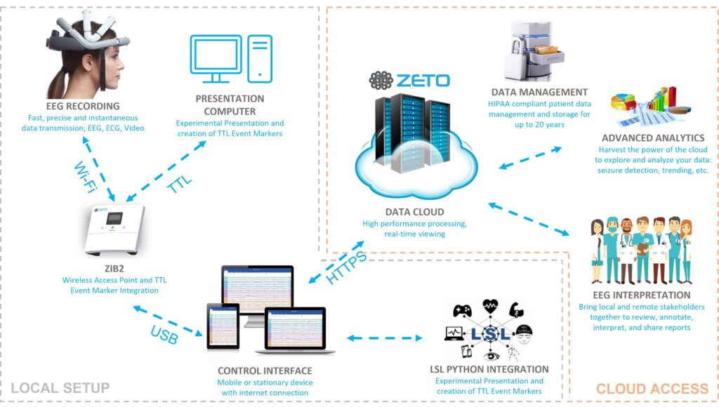
With Zeto’s event marker integration, users benefit from accurate event marker timing and distributed cloud data access and management – see Figure 1.

Figure 1. Schematic diagram of Zeto’s local event marker integration and remote data streaming and management features. Event markers from the presentation computer timing are merged with the EEG data locally via the Zeto Interface Box 2 (ZIB2) and then passed on to the Zeto Cloud. A simultaneous LSL integration and related multi-model data recording become possible out of the box while maintaining Zeto’s existing cloud streaming, data, and user management features.
ZETO ERP FEATURES
The Zeto EEG platform offers the ability to integrate external markers wirelessly at an 8-bit resolution within a 2 ms delay and less than 1 ms technical jitter. In other words, the user can distinguish between 255 unique event markers that they can repeat as closely as 4 ms from one another. With these features, Zeto EEG is equipped to reveal accurate auditory, visual, and senso-motoric ERPs across a wide range of applications.
Event markers are collected by the system via an 8-bit DB9 connector built into the Zeto Interface Box Version 2 (ZIB2) using Transistor-Transistor-Logic (TTL) signals.6 The ZIB2 acts as a data access point for the wireless Zeto WR19 headset. Synchronization between the ZIB2 and Zeto WR19 is handled at a nano-second range, eliminating both the delay and jitter introduced by the wireless data transmission. Incoming event markers are retroactively aligned with the data point at the time of collection.
Users can extract the event marker data from the Zeto system in multiple ways:
1) EDF+ File
Zeto users can export finished EEG recordings in various ways but the most popular is the EDF+ file format that is readable by most common EEG analysis tools. Event markers appear in the EDF+ file as digital I/O channels synchronized with the EEG data. Some EDF readers will also display the embedded event markers in the viewer.
2) Visualization
In the Zeto cloud software, users can visualize the TTL event marker inputs along with the EEG by selecting the “ALL” montage in the montage menu. This view is particularly useful for troubleshooting when setting up the ERP experiment. Offline or in real-time the user will see incoming event marker codes visualized high or low values in separate channels. The event marker channels are simultaneously translated into event marker labels that co-appear at the bottom of the screen (Figure 2).

Figure 2. Close-Up of the “All” Display montage: Output (“A”) or Input (“B”) event marker channels for 8 bits each, translated into up to 8-bit (255) unique event marker labels. The event marker mapping can be freely configured and labeled as desired prior to the recording. In this example, eight input event marker signals are embedded in the data file (pins 1 to 8) and show up as square waves (“C”). Each input event marker channel is mapped to an annotation, labeled “One” through “Eight” respectively at the bottom of the data screen.
3) Real-time lab streaming layer (LSL) Export
Eight digital input and output channels each are made available via lab streaming layer (LSL) API in real-time, enabling the user to note the stimulus onset directly in the data stream. Event makers and EEG are synchronized and merged prior to providing this data to the LSL streaming socket. As a result, event timing and EEG data remain perfectly synchronized even if there are LSL related streaming delays. Additional LSL API synchronization features remain available to users for additional real-time data integration.
4) Offline Event Marker Files
Users have the option to export marker files after the recording is completed. That marker file contains precise marker timing and label information for all event markers recorded for easy processing in third party analysis tools such as MATLAB, ERPLAB, Python or others. This allows for separate analysis of event data and EEG data found in the exported EDF+ file.
The event file can be exported in “.csv” format (Figure 3), or a comma-delimited format called “.zmrk”. Both are compatible with most common EEG processing tools currently available for research.

Figure 3. Event Markers listed in .csv format.
ZETO EVENT MARKER TIMING VALIDATION
Zeto validated the event marker timing to establish the delay and jitter attributes under working conditions.
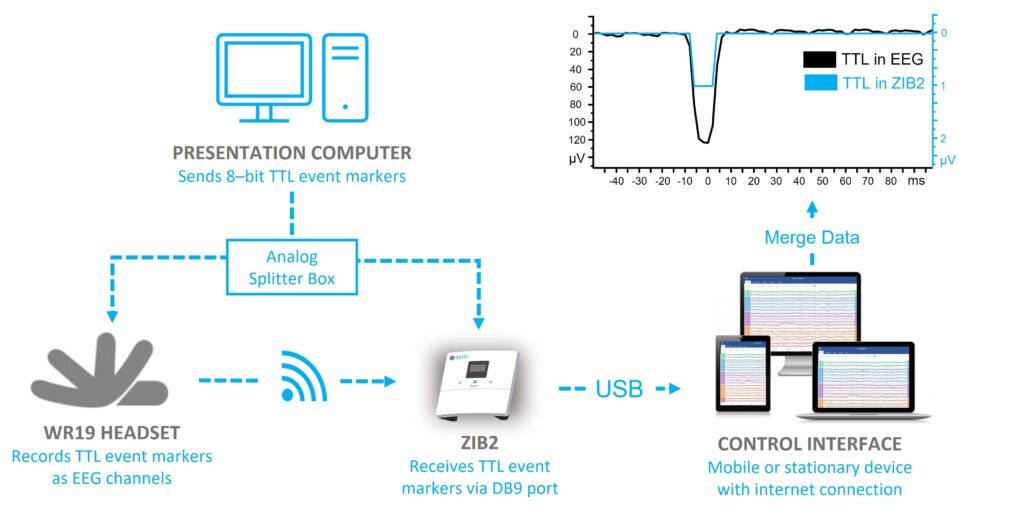
To do this, a testing setup split the incoming TTL trigger voltages via an analog splitter into two exact 8-channel copies. One copy of the event marker signals got connected to the ZIB2 input trigger ports while the second copy got connected to 8 channels of the WR19 headset. As a result, incoming event markers both appeared as digital events in the datastream and voltage changes in the EEG channels (Figure 4). Subsequent processing revealed the real-life delay and jitter between the incoming event marker signals and the EEG recording.

Figure 4. Schematic of the event marker timing test setup. The presentation computer sends 8-bit TTL event markers to an analog splitter box. One copy of the TTL signals arrives at the ZIB2 and gets converted into event labels. The other copy arrives at the headset and feeds into 8 of the EEG channels to show up as signals in the EEG data file.
Using this approach, event marker timing was established as stable, at < 2 ms delay and <1 ms jitter, which is a good basis to reliably capture ERP signals in EEG.
ZETO’S STIMULATION AND SYNCHRONIZATION PLATFORM PARTNERS
It is important to note that the Zeto system provides extremely precise synchronization on the receiving end of the event marker only. In fact, a much more likely source of both delay and jitter in ERP experiments occurs during stimulus presentation and subsequent event marker generation.
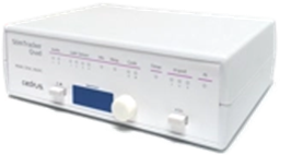
To avoid timing complications that occur prior to event markers entering the Zeto system, Zeto has partnered with two stimulus and synchronization platforms – both tested with our products. These stimulus presentation and synchronization products are designed to eliminate delay and jitter on the event marker onset. In addition, they offer a variety of additional functions, including experiment writing and presentation software, participant response boxes, and photodiodes.
Both partners have implemented out-of-the-box integrations for Zeto and are ready to service Zeto customers.


| Cedrus, Inc. | Psychology Software Tools |
| Cedrus devices are designed for precise, jitter-free event marking and fit a variety of budgets. SuperLab is an experiment writing application, while software support for their hardware interfaces also includes Matlab, E-Prime, Python, C++, etc. | Psychology Software Tools isa prominent software and hardware company that helps researchers address challenges in human behavioral studies. PST are creators of E-Prime, a market-leading experiment writing platform. |
 |  |
| C-POD Sent precise event markers via USB | Chronos Hardware synchronization and delivery system for millisecond-accurate event markers to external devices using various I/O port options. |
| M-POD Sent precise event markers, plus incorporate a response pad and photodiode | E-Prime Experiment writing platform that integrates stimulus presentation and behavioral software with research equipment |
| StimTracker Duo Comprehensive audio/visual/response synchronization platform | |
| SuperLab Easy to use experiment writing software that does not require programming |
References
- Hai Li et al. (2018). “Combining Movement-Related Cortical Potentials and Event-Related Desynchronization to Study Movement Preparation and Execution.” Frontiers in Neurology.
https://www.frontiersin.org/articles/10.3389/fneur.2018.00822/full - Fedor Jagla, Vladislav Zikmund, in Studies in Visual Information Processing (1994). “Visual and Oculomotor Functions.” ScienceDirect.
https://www.sciencedirect.com/topics/neuroscience/movement-related-potential - Sean McWeeny, Elizabeth S. Norton. “Understanding Event-Related Potentials (ERPs) in Clinical and Basic Language and Communication Disorders Research: A Tutorial.” PMC.
https://www.ncbi.nlm.nih.gov/pmc/articles/PMC3016705/ - Shravani Sur, V. K. Sinha (2009). “Event-related potential: An overview.” PMC.
https://www.ncbi.nlm.nih.gov/pmc/articles/PMC3016705/ - Thomas Hörberg et al. (2020). “Olfactory Influences on Visual Categorization: Behavioral and ERP Evidence.” Cerebral Cortex.
https://academic.oup.com/cercor/article/30/7/4220/5811850 - Fiorenzo Artoni et al. (2017). “Effective Synchronization of EEG and EMG for Mobile Brain/Body Imaging in Clinical Settings.” PMC.
https://www.ncbi.nlm.nih.gov/pmc/articles/PMC5770891/

