
Sweat artifacts are a common problem in electroencephalography (EEG) recordings. They can noticeably affect the quality of the recorded tracings and make it difficult to read the underlying EEG signals. Sweat artifacts in EEGs occur when the body’s biological sweat response alters the conductivity of the skin in a way that affects the electric signals picked up by the electrodes. Such changes not only occur when sweat is visible on the scalp but also occur when the body heats up and prepares to sweat.
In this blog, we will explore the causes of EEG sweat artifacts, their effects on EEG recordings, and strategies for mitigating their impact.
What Causes Sweating
Sweat is crucial for human thermoregulation and can be caused by a variety of factors, including anxiety, nervousness, or physical exertion. Biological changes during menopause also increase the chance of sweating. Regardless of these and other factors, a warm testing environment is the main driver of sweating.1 Systematic studies revealed that temperatures above 79°F (~26°C) can have a noticeable effect on the EEG and signal morphology.2
The Biology of Sweat Artifacts in EEG
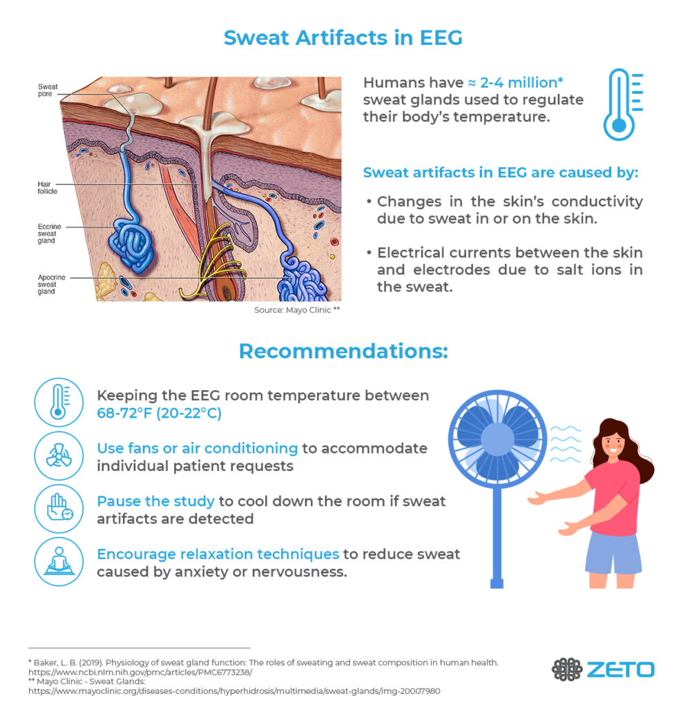
Sweating is not simply the appearance of sweat on the skin but the result of a cascade of biological changes that lead to the skin’s ability to secrete liquid from the sweat glands, onto the skin (Figure 1).
The filling of the sweat glands with liquid in preparation of sweat excretion increases the electrical conductivity of the skin rapidly which affects the morphology of the EEG signals. These rapid changes in skin conductivity and the uneven distribution of the sweat glands across the skin result in recordings prone to major EEG artifacts, with single channels showing large signal changes at different times and locations.3
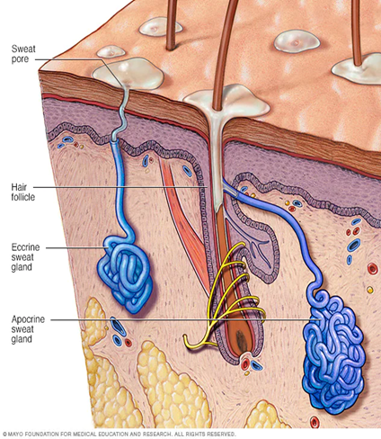
Figure 1. Cross section of epidermis and dermis skin layers with embedded hair follicle, eccrine, and apocrine sweat glands. Source: Mayo Clinic
The Physics of Sweat Potentials in EEG
In addition to biological changes in the skin’s conductivity, the composition of the sweat itself is contributing to electrical potentials that EEG amplifiers pick up. Sweat contains high sodium chloride and lactic acid which react with metallic components of the EEG electrodes, generating electrical potentials.4 These electrical potentials combine with skin and sweat gland potentials into what is visible in the EEG as sweat artifacts.
Appearance of Sweat Artifacts in EEG
Sweat artifacts in EEG can appear in various morphologies or shapes that are affected by biological factors such as the severity and generality of the sweat response. The sudden onset of sweating across the entire body will appear different from sweating that occurs over time or may be more limited by body part or region. More relevant for the appearance in EEG though, are the analog or digital filter settings of the recording.
Amplifiers with a built-in low-frequency hardware filter will show a more subdued sweat artifact even without any digital filtering. True direct current (DC) amplifiers that do not have any analog low-cut-off filter will show the build-up to a sweat artifact in their raw data much more because small changes over time can be picked up much better.
Most clinical EEGs are viewed at a 1 Hz–70Hz bandpass filter as recommended by ACNS.5 EEG Sweat artifacts viewed using a 1 Hz low-cut-off filter generally show up as slow wave components around a 1 Hz–3 Hz frequency in otherwise normal background activity; for an example, see Figure 2. Disabling the low-cut-off filters, however, exposes additional low-frequency drifts related to sweat that are otherwise masked by digital signal processing; for an example, see Figure 3.
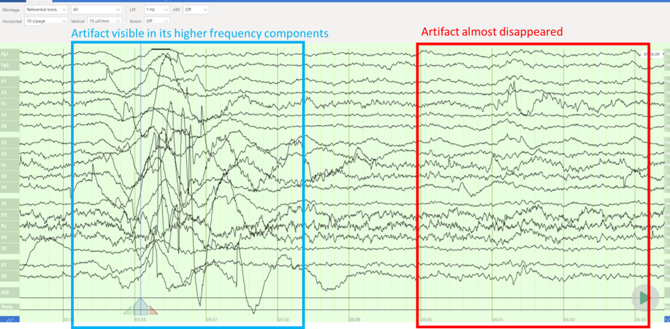
Figure 2. Filtered sweat artifact in a full 19-channel clinical EEG viewed in a referential montage. 1 Hz low-frequency forward Butterworth filter applied. The slow meandering signal drifts almost completely disappears after filtering (red frame).
Sharper signal drifts remain visible even after filtering (blue frame). For most clinical recordings, EEG tracings such as this are indicators of the biological changes that are caused by a sweat response. Data was recorded using Zeto’s WR19 headset at 79°F (~26°C).
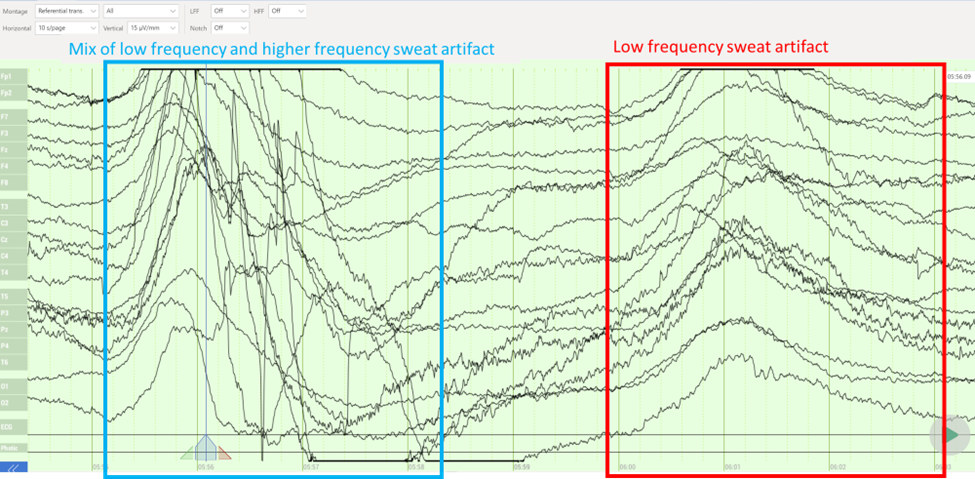
Figure 3. Unfiltered sweat artifact during the same data segment, as presented in Figure 2. Slow meandering (red frame) and at times sharper signal drifts (blue frame) reflect the biological changes in the skin’s conductivity due to sweating.
How to Get Rid of Sweat Artifacts in EEG
There are two common ways to reduce or avoid sweat artifacts in EEG recordings.
- EEG operators can reduce the biologically triggered changes that lead to sweating. In preparation for the EEG recording, operators can ask patients to avoid strenuous exercise, caffeine, and alcohol prior to a scheduled EEG session, ideally 24 hours before the test. During EEG recordings, Kappenman and Luck recommend maintaining a cool temperature in the recording environment to minimize the occurrence of EEG sweat artifacts. They recommend a comfortable temperature of 68°F –72°F (20°C –22°C) and using fans or air conditioning to prevent humidity buildup.2
- During the EEG session, EEG operators should assure the best possible electrode contact with the scalp to reduce skin impedance under the electrode. In traditional amplifier systems with wired electrodes, this can be achieved via additional skin preparation and abrasion. With active quick-apply EEG recording systems, such as Zeto’s headset, operators can assure proper electrode landing with each conductive leg touching the scalp.
Bottom Line – Recommendations
In hectic clinical day-to-day EEG schedules, the easiest way to avoid sweat artifacts in most patients is to avoid sweating in the first place. For that reason, option #1, mentioned previously (reducing sweating), is the most robust way to assure consistent EEG data quality.
- Keep the EEG room temperature at 68°F – 72°F (20°C–22°C), especially when recording unconscious patients who cannot communicate their comfort levels; keeping an optimal temperature reduces the body’s need for sweating.
- Use fans or air conditioning to accommodate individual patient’s temperature requests. Each patient is different; ask to make sure they are not hot.
- If EEG sweat artifacts are detected, consider pausing the study to cool down the room (i.e., opening the door, reducing the room temperature, and/or the use of a fan).
- Relaxation techniques: Encouraging the patient to relax and breathe deeply. This can help to reduce sweat caused by anxiety or nervousness.
By implementing these strategies, EEG operators can help minimize sweat artifacts in EEGs and obtain cleaner results. It is important to work closely with the patient and monitor the EEG tracings for any signs of EEG sweat artifacts during the test to more adequately address issues with data quality.
References
- Baker, L. B. (2019). Physiology of sweat gland function: The roles of sweating and sweat composition in human health.
https://www.ncbi.nlm.nih.gov/pmc/articles/PMC6773238/ - Kappenman, E. S., & Luck S. J. (2010). The effects of electrode impedance on data quality and statistical significance in ERP recordings.
https://static1.squarespace.com/static/5abefa62d274cb16de90e935/t/5ac6962a8a922d0b8b8be6a1/1522964012664/Kappenman+2010+Psychophys+Impedance.pdf - Kalevo, L., Miettinen, T., Leino, A., Kainulainen, S., Korkalainen, H., Myllymaa, K., … & Myllymaa, S. (2020). Effect of sweating on electrode-skin contact impedances and artifacts in EEG recordings with various screen-printed Ag/Agcl electrodes.
https://ieeexplore.ieee.org/stamp/stamp.jsp?arnumber=9017959 - Siddiqui, F., Osuna, E., Walters, A., Chokroverty, S. (2006). Sweat artifact and respiratory artifact occurring simultaneously in polysomnogram.
https://pubmed.ncbi.nlm.nih.gov/16461004/ - ACNS – American Clinical Neurophysiology Society Guidelines and Consensus Statements.
Guidelines and Consensus Statements | ACNS – American Clinical Neurophysiology Society