
Video EEG is an important tool in diagnosing and monitoring patients with epilepsy or other neurological conditions. It helps distinguish physiologic or external artifacts from epileptic seizures or epileptiform discharges associated with seizures. Video EEG also helps to differentiate epileptic seizures from psychogenic nonepileptic spells that are mistaken for seizures and other episodic abnormal movements associated with other conditions, such as tic disorders, tremor, or periodic limb movements of sleep. Simultaneous video and EEG recordings enable the establishment of correlations between abnormal movements and abnormal wave activity.1,2,3,4
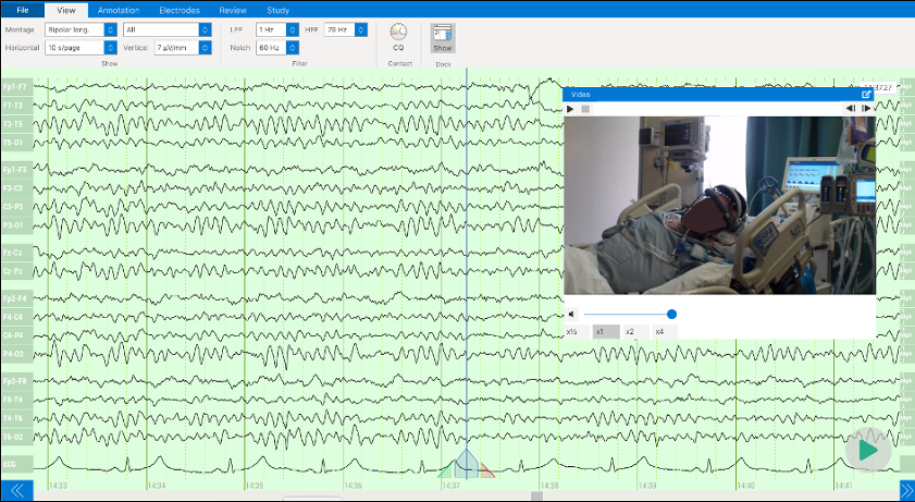
For awake patients, video can identify sources of noise such as muscle artifacts, blinking, chewing, forehead wrinkling, and even ear wiggling. For unconscious patients, video can identify external sources of artifact, such as electronic devices in the ICU (for example, an artificial respiration device). The inclusion of video in the EEG improves the ability to accurately diagnose a patient. Additionally, video can confirm correct electrode placement and headset positioning.
In this blog, we discuss the benefits of using video integration in EEG and what Zeto EEG can offer.
Different noise sources in EEG
EEG recordings can be impacted by various sources of noise and interference, including physiological noise, environmental noise, EEG electrode placement, electrical artifacts from EEG electrode malfunctions or from external electrical devices, and motion artifacts. These artifacts can make it difficult to distinguish genuine brain activity from noise, compromising the ability to accurately interpret EEG recordings.
Physiological noise arises from the body’s internal processes like muscle contractions, heartbeats, and eye movements, generating electrical signals that can interfere with the EEG recording. Environmental noise, on the other hand, encompasses external disturbances like electrical noise from equipment, electromagnetic interference, or ambient noise, which can introduce unwanted signals into the EEG data. Electrical artifacts can also be produced by the EEG equipment itself, resulting from issues like poor grounding, incorrect amplifier settings, or malfunctioning electrodes. Lastly, motion artifacts arise from bodily movements such as head motions, jaw opening and closing, or eye blinks, leading to distortions in the EEG signal and complicating accurate data interpretation.
To account for artifacts and enhance the interpretation of EEG recordings, video recording is often used in conjunction with EEG to identify and exclude sources of noise from the data.
Video EEG: Complement or Necessity?
Video is particularly useful for identifying artifacts related to movements or other physical activities that may produce electrical signals in the EEG recording. For example, if a patient moves their arm during an EEG recording, this movement can cause changes in the electrical activity picked up by the electrodes, which is recorded in the EEG. By observing the video recording, it is possible to identify the movement as an artifact and avoid misinterpreting the EEG data. This helps ensure that EEG signals identified as epileptiform activity represent genuine brain activity, rather than artifacts from other sources. Epileptiform waves can sometimes be difficult to distinguish from artifacts, as they can have similar characteristics in the EEG recording.
Video can also help diagnose and classify epileptic seizures and distinguish them from psychogenic nonepileptic spells and episodic abnormal movements due to other medical conditions that are not epileptic seizures.1,2,3,4,5 For example, if a patient experiences an epileptic seizure during an EEG recording, the video can help correlate the EEG signal with the visible movements and behaviors of the seizure. A psychogenic nonepileptic spell will show motion artifacts on the EEG but will not show underlying abnormal brain wave activity. Furthermore, certain behaviors such as squeezing the eyes shut while shaking, awareness during the seizure, and returning to baseline mental status right after the event can suggest a psychogenic cause.5
Sometimes tics, tremors, muscle twitching, and other abnormal movements in the tongue or a limb or digit can be mistaken for epileptic seizures, particularly in the ICU setting where subclinical seizures occur more frequently. Subclinical seizures are epileptic seizures that are not associated with characteristic abnormal movements usually associated with seizures. Video EEG can determine if the suspicious subtle abnormal movements are in fact due to epileptic seizures in most cases. If the abnormal movements represent subclinical epileptic seizures, they would be expected to correlate with epileptic seizure brain wave activity on the EEG.
Video recording provides additional information about the patient’s behavior or movements at the time of a seizure or seizure-like event, which is called a seizure’s semiology.1,3,5 Gaze deviation and arm stretching right before the seizure or at seizure onset can point to the seizure origin in the brain and inform localization of seizure foci relative to brain hemispheres or specific areas such as the motor cortex, supplementary motor areas, and frontal and parietal eye fields. Seizure semiology also includes the subjective sensations and experiences immediately preceding and during a seizure. It is critical to the diagnosis and classification of seizure disorders, which in turn affects the treatment plan. A video EEG is critical to presurgical evaluation prior to epilepsy surgery to diagnose and classify epileptic seizures when present and avoid unnecessary surgery due to psychogenic nonepileptic spells or other episodic abnormal movements not due to epileptic seizures.1
Zeto EEG: Seamless Compatibility with Any Camera, Reimbursable, Supports Up to Four Cameras
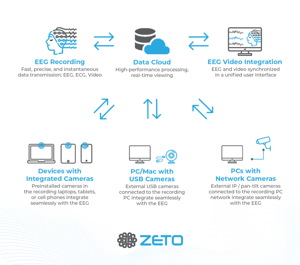
Zeto’s video integration feature reshapes the users expectations of what is possible with modern technology. The Zeto system enables simple camera controls (zooming and panning) through the cloud, ensuring simple usability.
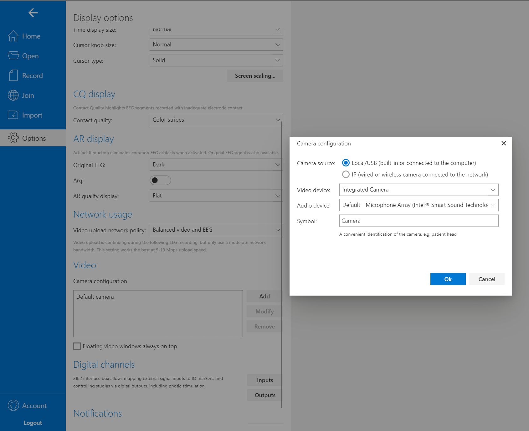
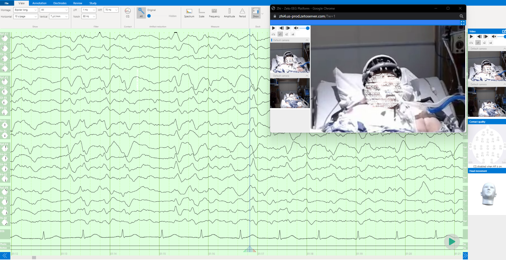
One of the most appreciated features by users of the Zeto platform is its compatibility with any integrated or USB enabled camera. That eliminates the need for additional camera purchases and helps operators to use Zeto EEG more easily across a wide range of settings and recording locations. The Zeto Cloud Platform supports the use of up to four cameras simultaneously, providing medical professionals with the flexibility to monitor patients from multiple angles.
In addition to its compatibility with integrated and external USB cameras, Zeto offers the simple integration of IP cameras, whether they are wired or wireless. This capability is particularly beneficial when medical personnel already have pre-installed IP cameras in the room, as they can add these cameras to the Zeto Cloud Platform for a seamless integration. IP camera integration often depends on existing local IT infrastructure and cybersecurity guidelines which can result in longer integration timelines compared to integrated or USB – based camera solutions. But with the added configuration time come additional conveniences and features (such as lowlight or night vision capabilities) which often make this extended setup more than worth it.
| Setup Time | Price | Ability to Point Toward Patient | Optical Zoom | Low Light / Night Vision | |
| Integrated Cameras | Near instantaneous | Integrated in most devices | Static angles – requires adjusting recording device | Mostly Unavailable | Mostly unavailable in built-in devices |
| External USB Cameras | Fast – plug-in and use | < $200 for most, <$400 for high-end | Manually able to point in any direction | Manual zoom in some devices | Available for additional cost |
| IP Cameras | Often requires IT support and additional setup | < $1,500 for most | Manually able to point or use of remote pan/tilt features | Automatic or manual zoom options available | Available in most devices |
With any of these camera options, maximum image resolution depends on the utilized camera hardware, but integrated, USB, or IP camera options are available with Ultra 4k resolutions or higher. The consideration then becomes data storage size and related storage costs more than the technical ability – providers may still choose to save data in lower resolution settings simply to save storage costs.
Another powerful tool of the Zeto Cloud platform is the ability for the clinician to remotely access the recording through any device connected to the internet. Allowing for real time analysis when the provider is not present in the recording environment.
Medical practitioners will find the real-time monitoring capability invaluable, enabling them to observe patients with precision. Additionally, Zeto’s video EEGs, utilizing the integrated cameras, are eligible for reimbursement through CPT codes, specifically for EEGs lasting 24 hours or longer.

By utilizing video, Zeto ensures accurate positioning of the headset and proper electrode placement. This attention to detail guarantees the collection of reliable and precise EEG data.
In conclusion, video recording is an important complement to EEG recordings. It helps identify and exclude sources of artifact in EEG and confirms the presence of epileptic seizure activity and epileptiform discharges important for the diagnosis and classification of epileptic seizures. Video EEG also helps to identify psychogenic nonepileptic events and episodic abnormal movements from other medical conditions mistaken for epileptic seizures. By using video in conjunction with EEG, clinicians can improve the interpretation of EEG recordings, leading to more accurate diagnoses and better treatment outcomes for patients with neurological conditions.
Zeto’s video integration feature enhances convenience and accuracy, ultimately resulting in better diagnosis and treatment outcomes for patients.
Sources:
- https://onlinelibrary.wiley.com/doi/10.1111/j.1528-1167.2006.00656.x
- https://www.ncbi.nlm.nih.gov/pmc/articles/PMC9259997/
- https://researchopenworld.com/wp-content/uploads/2020/06/AWHC-3-3-315.pdf
- https://www.sciencedirect.com/science/article/abs/pii/0887899495000217
- https://www.ncbi.nlm.nih.gov/pmc/articles/PMC4206377/